A Patient's Guide to the Normal Ear
The Anatomy and Function of the Normal Ear
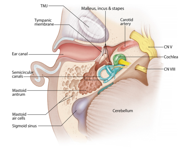
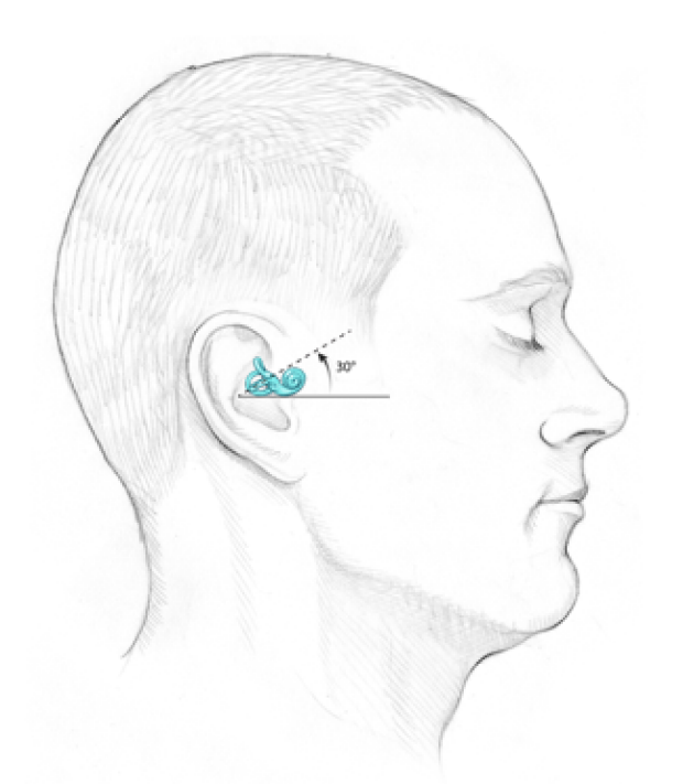
The ear consists of the organs of hearing and balance. These are located within the temporal bone in the base of the skull.

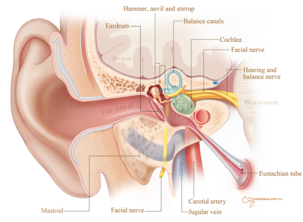
The external ear includes the visible part of the ear (the auricle) and the ear canal. This system allows the air vibrations of sounds to pass from the environment to the ear drum.
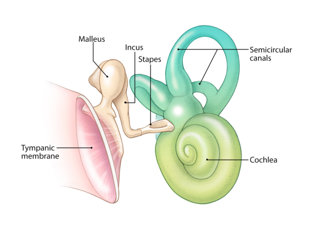
The middle ear includes the eardrum (tympanic membrane) and the air-filled chamber behind it. The middle ear space contains the three bones of hearing, the malleus (“hammer”), incus (“anvil”) and stapes (“stirrup”). In the normal ear, the tympanic membrane vibrates from sound, the three bones (or ossicles) move as a unit to transmit the sound energy to the inner ear. The middle ear space is connected to the back of the nose through the Eustachian tube, this allows air to enter the middle ear and equalize pressures with the outside environment.
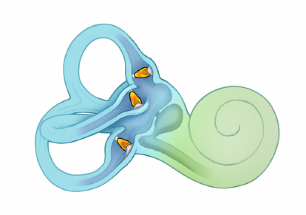
The inner ear consists of the cochlea (organ of hearing), the vestibular system (organ of balance), and the nerves that travel to the brain. The cochlea converts mechanical sound vibrations to electrical nerve impulses. The vestibular system is made up of the balance canals which sense rotation, and the otolith organs which sense linear motion.

The Anatomy and Function of Normal Hearing
Sound is a vibration that enters the ear though the ear canal. This moves the tympanic membrane (“eardrum”) and the three small bones of hearing in the middle ear (malleus, incus, and stapes).
The stapes vibrates as a piston against the fluid of the hearing postion of the inner ear (cochlea).
The cochlea converts these mechanical sound vibrations to electrical nerve impulses. There are fragile membranes that divide the cochlea into 3 fluid-filled spaces (or scalae) through its 2.5 turns. The stapes attaches to the inner ear at the oval window, where transmits the sound vibrations into the scala vestibuli. The vibration travels all the way up the 2.5 turns in the scala vestibuli tho the apex of the cochlea, where it connects with the scala vestibuli. At this point, the vibration travels all the way back down the the scala tympani, back through the 2.5 turns to be released into the middle ear through a membrane called the round window.

The movement of the fluids within the cochlea from these traveling vibrations moves the thin membrane (basilar membrane) that holds the Organ of Corti. The normal organ of Corti contains about 16 thousand microscopic hair cells. The hairs on these cells move as a result of the sound vibrations, and this causes an electrical signal to be passed to the nerves in the core of the cochlea. The electrical signal is passed through these nerves to the brain where it is perceived as sounds.

An important part of cochlea’s function is its “tonopopic organization”. This means that different sound frequencies stimulate different parts of the cochlea. So, high frequencies stimulate the base (closest to the stapes) more, while low frequencies stimulate the apex (farthest from the stapes) more.
The nerves that attach to the hair cells at various locations are therefore “tuned” to be more sensistive to specific frequencies.
In this way, the inner ear begins to process complex sound signals by breaking it down into various frequencies.
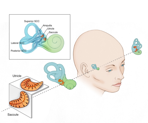
The Anatomy and Function of Normal Balance
The inner ear is the organ of balance (vestibular system) that provides us with information when we move. There are two major parts to the vestibular system, the semicircular canals (that sense angular acceleration) and the otolith organs (that sense linear aceleration).
Semicircular canals
Each inner ear contains 3 balace canals (semicircular canals). These are ear oriented at 90 degrees to each other.
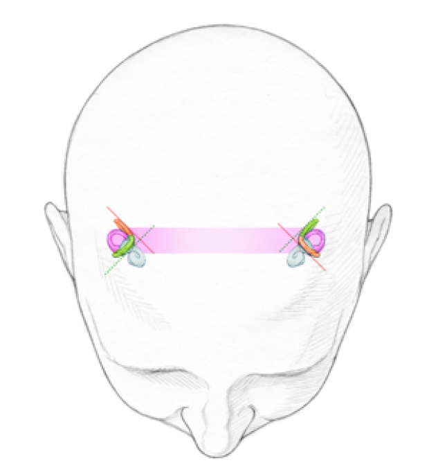
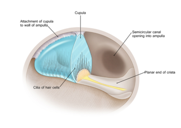
When the head moves in the plane of one of the canals, the fluid in the canals moves relative to the inner ear. This deflects a membrane (cupula) in one end of the canal (the ampulla). The membrane motion deflects the microscopic hairs in the ampulla, which in turn send an electrical signal to the brain though the balance nerves.


Because the semicircular canals are all oriented in different planes, each specifically senses turning motion (angular acceleration) in a particular direction.
Otolith organs
The otolith organs (utricle and saccule) provide information on movement in a straight line and tipping (linear acceleration). These organs are located in the vestibule portion of the inner ear. They are each made up of many hair cells arranged in a sheet. The ends of these hair cells are embedded in a gelatin layer that contains small calcium crystals. When we move or tip our heads, the calcium crystals move as well, deflecting the hairs in the otolith organs. This deflection stimulates the balance nerves, tells us when we move, and which direction is up (linear acceleration).