Multidisciplinary Head & Neck Cancer Care
Head & Neck Surgery
We are the pioneers of major scientific breakthroughs
- Organ preservation approaches to head and neck cancer.
- New drugs for head and neck squamous cell carcinoma (HNSCC) and extending uses of existing drugs to HNSCC and nasopharyngeal carcinoma (NPC).
- Advanced radiation therapy techniques that limit toxicity and improve outcomes.
- Minimally Invasive and Robotic Surgery
- Stem cell work that extends the findings of the first paper, demonstrating the existence of “cancer stem cells” in HNSCC by researchers from Stanford and Michigan in 2007; and a 2009 Stanford study establishing that stem cell properties of patients’ malignancies correlate with prognosis. This work led to subsequent stem cell papers in 2011 and 2012.
- Normal tissue stem cell studies to identify salivary gland stem cells and to manipulate them for preservation and/or restoration of salivary gland function from radiation damage.
- HNOP’s breadth of research studies and protocols including treatment of intermediate and advanced disease as well as hypoxia imaging.
Our Innovations
- Creation of the first head and neck multidisciplinary tumor patient conference (tumor board; 1976) in the U.S.
- Introduction of the first use of chemotherapy with irradiation for head and neck squamous cell carcinoma (HNSCC), which is the basis of organ-preservation chemoradiation in the U.S.
- Close working relationships with:
- Neurosurgery, Interventional Radiology, and Neuroradiology,which are critical for complex open and endonasal endoscopic skull base surgery.
- Endocrinology in the treatment of thyroid cancer.
- Dermatology in the treatment of advanced skin cancers.
- Innovative research by physicians now at Stanford that demonstrates the utility of the FDA-approved Mobetron for intraoperative radiation therapy.
- Contributing research in a Phase II trial of immunotherapy in intermediate and advanced surgically-treated HNSCC. A Phase III trial is now planned.
- Leadership in the head and neck disease site committee of the Radiation Therapy Oncology Group to develop new nation-wide clinical trials in head and neck cancer.
- Biomarker studies to identify novel circulating biomarkers for prognostication and post-treatment surveillance in head and neck cancer.
- Strong links to developmental therapeutics such as the advancement of new drugs to treat cancer.
- Provision of a full range of treatment options that include minimally invasive surgery, robotic surgery, stereotactic radiosurgery such as CyberKnife, microvascular reconstruction, intraoperative radiation therapy (IORT), and new chemotherapy trials.
What is Head & Neck Cancer?
Head and neck cancer is a term that can include the broad array of tumors which may arise in this anatomically diverse region of the human body. Most often, the term head and neck cancer refers to tumors that arise from “squamous” cells that line the moist, mucosal surfaces of the mouth and throat. In fact, 95% of head and neck tumors are squamous cell carcinoma.
Tumors of the thyroid, salivary, and parathyroid glands, as well as cancers of the brain, nose and paranasal sinuses, esophagus, and eye, are not usually categorized as head and neck cancer. Furthermore, tumors of the skin, muscle and bone arising in the head and neck are also typically not included in this term.
Head and neck cancer is then further classified by its location within the mouth and throat:
Oral cavity
The lips, the oral tongue” (the forward two-thirds or front part of the tongue), the gums lining the upper and lower jaws, as well as the lining inside the cheek. The area known as the floor of the mouth is a mobile area between the lower jaw and gum and the oral tongue. The roof of the mouth or “hard palate” is also included as part of the oral cavity. Finally, a small triangulated area of mucosa or gum lining the area behind the last wisdom tooth is called the“retromolar trigone” and is also part of the oral cavity.
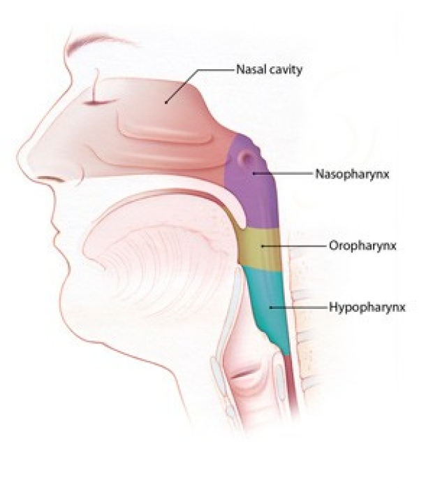
Pharynx
In medical terminology, the throat is known as the pharynx. In fact, the pharynx is supple tube or funnel that connects both the nose and mouth to the swallowing tube or esophagus. The pharynx is composed of three parts: the nasopharynx (the area just behind the nose); the oropharynx (behind the oral cavity and in the back of the mouth], and the hypopharynx, which surrounds the voice box and leads into the esophagus.
The larynx critical not only for the production of speech, but also breathing and swallowing. The “supraglottic” larynx has a valve called the epiglottis, which covers the larynx during swallowing to prevent “aspiration” of food into the lungs.

Treatments
HNOP offers multi-disciplinary, collaborative and integrated evaluation and care for patients with head and neck cancers.
Minimally invasive or endoscopic head and neck surgery (eHNS) is a dynamic new approach that allows surgeons to remove tumors with use of a specialized endocopes and cameras without external incisions and usually with little or no change in speech, appearance, and swallowing function.
An endoscope is a long, thin tube with special lighting and a narrow lens through which the surgeon can view organs and tissue inside of the body. For throat cancers, the surgeon inserts the endoscope through the patient’s mouth, and a microscope provides an excellent image of the tumor. Using very precise, state-of-the-art surgical instruments that are also inserted through the mouth, the surgeon can perform the operation without an external incision.
eHNS has several advantages in many cases. Some of these advantages are:
- Reduced risk of blood loss
- Lower pain levels
- Fewer days spent in the hospital
- Quicker return to a normal diet and faster recovery time
- Less scarring, with improved cosmetic appearance
In some cases of throat cancer, eHNS may reduce or even eliminate the need for chemotherapy and radiation therapy.
At Stanford, your team of surgeons, oncologists, and radiologists will work together to determine the best course of action for you. The goal with eHNS is always the same: to eliminate the cancer while minimizing the risks and recovery time associated with traditional cancer care.
Chemotherapy or radiation therapy may still be necessary after eHNS. When chemotherapy and radiation therapy cannot be avoided through surgery, eHNS may still hold advantages for patients. Faster recovery after eHNS means patients can usually begin chemotherapy and radiation therapy earlier. And the smaller incisions with eHNS heal more quickly than larger incisions and are less likely to become infected.
Two types of eHNS, transoral robotic surgery (TORS) and transoral laser CO2microsurgery (TLM), have revolutionized the treatment for throat cancer.
Robotic surgery uses state-of-the-art technology that allows surgeons to safely remove certain thyroid tumors through discrete incisions several inches from the neck. Because no incision is made in the neck, the patient avoids a neck scar.
The da Vinci ® surgical system is a highly sophisticated computerized system that is used for robotic thyroid surgery. The surgeon cuts a 1-inch to 2-inch incision in the folds of skin under the patient’s arm and inserts the da Vinci ® system’s robotic arms, which have been customized to resemble standard surgical instruments. The surgeon guides the robotic arms underneath the skin toward the thyroid gland.
The surgeon views the surgical field on a 3-D high-definition screen, magnified up to 10 times. The skilled surgeons at Stanford Medicine control the da Vinci ® system’s robotic arms as one might control conventional surgery. The instruments on the robotic arms can move with seven degrees of movement and rotate 540 degrees, giving the surgeon the ability to manipulate delicate tissues with precision.
Robotic thyroidectomy is not available for all thyroid tumors. If your thyroid tumor is less than 3 cm in size and is likely to be benign (non-cancerous), then robotic surgery may be an option. Your surgeons will help you decide the best approach for you.
Testing Docetaxel-Cetuximab or the Addition of an Immunotherapy Drug, Atezolizumab, to the Usual Chemotherapy and Radiation Therapy in High-Risk Head and Neck Cancer
This phase II/III trial studies how well radiation therapy works when given together with cisplatin, docetaxel, cetuximab, and/or atezolizumab after surgery in treating patients with high-risk stage III-IV head and neck cancer the begins in the thin, flat cells (squamous cell). Specialized radiation therapy that delivers a high dose of radiation directly to the tumor may kill more tumor cells and cause less damage to normal tissue. Drugs used in chemotherapy, such as cisplatin and docetaxel, work in different ways to stop the growth of tumor cells, either by killing the cells or by stopping them from dividing. Cetuximab is a monoclonal antibody that may interfere with the ability of tumor cells to grow and spread. Immunotherapy with monoclonal antibodies, such as atezolizumab, may help the body's immune system attack the cancer, and may interfere with the ability of tumor cells to grow and spread. The purpose of this study is to compare the usual treatment (radiation therapy with cisplatin chemotherapy) to using radiation therapy with docetaxel and cetuximab chemotherapy, and using the usual treatment plus an immunotherapy drug, atezolizumab.
Stanford is currently accepting patients for this trial.
Stanford Investigator(s):
Intervention(s):
- radiation: intensity-modulated radiation therapy
- drug: cisplatin
- drug: docetaxel
- biological: cetuximab
- other: laboratory biomarker analysis
- drug: Atezolizumab
- other: Quality-of-Life Assessment
- procedure: Biospecimen Collection
- procedure: Computed Tomography
- procedure: Magnetic Resonance Imaging
- procedure: Biospecimen Collection
- procedure: Computed Tomography
- procedure: Biospecimen Collection
- procedure: Computed Tomography
- procedure: Magnetic Resonance Imaging
- procedure: Magnetic Resonance Imaging
- procedure: Biospecimen Collection
- procedure: Computed Tomography
- procedure: Magnetic Resonance Imaging
- procedure: Biospecimen Collection
- procedure: Computed Tomography
- procedure: Magnetic Resonance Imaging
- procedure: Biospecimen Collection
- procedure: Computed Tomography
- procedure: Magnetic Resonance Imaging
- procedure: Biospecimen Collection
- procedure: Computed Tomography
- procedure: Magnetic Resonance Imaging
- procedure: Biospecimen Collection
- procedure: Computed Tomography
- procedure: Magnetic Resonance Imaging
- procedure: Biospecimen Collection
- procedure: Computed Tomography
- procedure: Magnetic Resonance Imaging
- procedure: Magnetic Resonance Imaging
- procedure: Biospecimen Collection
- procedure: Computed Tomography
- procedure: Magnetic Resonance Imaging
- procedure: Biospecimen Collection
- procedure: Computed Tomography
- procedure: Magnetic Resonance Imaging
- procedure: Biospecimen Collection
- procedure: Computed Tomography
- procedure: Magnetic Resonance Imaging
- procedure: Biospecimen Collection
- procedure: Computed Tomography
- procedure: Magnetic Resonance Imaging
- procedure: Biospecimen Collection
- procedure: Computed Tomography
- procedure: Biospecimen Collection
- procedure: Computed Tomography
- procedure: Magnetic Resonance Imaging
- procedure: Biospecimen Collection
- procedure: Computed Tomography
- procedure: Magnetic Resonance Imaging
- procedure: Biospecimen Collection
- procedure: Computed Tomography
- procedure: Magnetic Resonance Imaging
- procedure: Biospecimen Collection
- procedure: Computed Tomography
- procedure: Magnetic Resonance Imaging
- procedure: Biospecimen Collection
- procedure: Computed Tomography
- procedure: Magnetic Resonance Imaging
- procedure: Biospecimen Collection
- procedure: Computed Tomography
- procedure: Magnetic Resonance Imaging
- procedure: Biospecimen Collection
- procedure: Computed Tomography
- procedure: Magnetic Resonance Imaging
- procedure: Biospecimen Collection
- procedure: Computed Tomography
- procedure: Magnetic Resonance Imaging
- procedure: Biospecimen Collection
- procedure: Computed Tomography
- procedure: Magnetic Resonance Imaging
- procedure: Biospecimen Collection
- procedure: Computed Tomography
- procedure: Magnetic Resonance Imaging
- procedure: Magnetic Resonance Imaging
- procedure: Biospecimen Collection
- procedure: Computed Tomography
- procedure: Magnetic Resonance Imaging
- procedure: Biospecimen Collection
- procedure: Computed Tomography
- procedure: Magnetic Resonance Imaging
- procedure: Biospecimen Collection
- procedure: Computed Tomography
- procedure: Magnetic Resonance Imaging
- procedure: Biospecimen Collection
- procedure: Computed Tomography
- procedure: Magnetic Resonance Imaging
- procedure: Biospecimen Collection
- procedure: Computed Tomography
- procedure: Magnetic Resonance Imaging
- procedure: Biospecimen Collection
- procedure: Computed Tomography
- procedure: Magnetic Resonance Imaging
- procedure: Biospecimen Collection
- procedure: Computed Tomography
- procedure: Magnetic Resonance Imaging
- procedure: Biospecimen Collection
- procedure: Computed Tomography
- procedure: Magnetic Resonance Imaging
- procedure: Biospecimen Collection
- procedure: Computed Tomography
- procedure: Magnetic Resonance Imaging
- procedure: Biospecimen Collection
- procedure: Computed Tomography
- procedure: Magnetic Resonance Imaging
- procedure: Biospecimen Collection
- procedure: Computed Tomography
- procedure: Magnetic Resonance Imaging
- procedure: Biospecimen Collection
- procedure: Computed Tomography
- procedure: Magnetic Resonance Imaging
- procedure: Biospecimen Collection
- procedure: Computed Tomography
- procedure: Magnetic Resonance Imaging
- procedure: Biospecimen Collection
- procedure: Computed Tomography
- procedure: Magnetic Resonance Imaging
- procedure: Biospecimen Collection
- procedure: Computed Tomography
- procedure: Magnetic Resonance Imaging
- procedure: Biospecimen Collection
- procedure: Computed Tomography
- procedure: Magnetic Resonance Imaging
- procedure: Biospecimen Collection
- procedure: Computed Tomography
- procedure: Magnetic Resonance Imaging
- procedure: Biospecimen Collection
- procedure: Computed Tomography
- procedure: Magnetic Resonance Imaging
- procedure: Biospecimen Collection
- procedure: Computed Tomography
- procedure: Magnetic Resonance Imaging
- procedure: Biospecimen Collection
- procedure: Computed Tomography
- procedure: Magnetic Resonance Imaging
- procedure: Biospecimen Collection
- procedure: Computed Tomography
- procedure: Magnetic Resonance Imaging
- procedure: Biospecimen Collection
- procedure: Computed Tomography
- procedure: Magnetic Resonance Imaging
- procedure: Biospecimen Collection
- procedure: Computed Tomography
- procedure: Magnetic Resonance Imaging
- procedure: Biospecimen Collection
- procedure: Computed Tomography
- procedure: Magnetic Resonance Imaging
- procedure: Biospecimen Collection
- procedure: Computed Tomography
- procedure: Magnetic Resonance Imaging
- procedure: Biospecimen Collection
- procedure: Computed Tomography
- procedure: Magnetic Resonance Imaging
- procedure: Biospecimen Collection
- procedure: Computed Tomography
- procedure: Magnetic Resonance Imaging
- procedure: Biospecimen Collection
- procedure: Computed Tomography
- procedure: Magnetic Resonance Imaging
- procedure: Biospecimen Collection
- procedure: Computed Tomography
- procedure: Magnetic Resonance Imaging
- procedure: Biospecimen Collection
- procedure: Computed Tomography
- procedure: Magnetic Resonance Imaging
- procedure: Biospecimen Collection
- procedure: Computed Tomography
- procedure: Magnetic Resonance Imaging
- procedure: Biospecimen Collection
- procedure: Computed Tomography
- procedure: Magnetic Resonance Imaging
- procedure: Biospecimen Collection
- procedure: Computed Tomography
- procedure: Magnetic Resonance Imaging
- procedure: Biospecimen Collection
- procedure: Computed Tomography
- procedure: Magnetic Resonance Imaging
- procedure: Biospecimen Collection
- procedure: Computed Tomography
- procedure: Magnetic Resonance Imaging
- procedure: Biospecimen Collection
- procedure: Computed Tomography
- procedure: Magnetic Resonance Imaging
- procedure: Biospecimen Collection
- procedure: Computed Tomography
- procedure: Magnetic Resonance Imaging
- procedure: Biospecimen Collection
- procedure: Computed Tomography
- procedure: Magnetic Resonance Imaging
- procedure: Biospecimen Collection
- procedure: Computed Tomography
- procedure: Magnetic Resonance Imaging
- procedure: Biospecimen Collection
- procedure: Computed Tomography
- procedure: Magnetic Resonance Imaging
- procedure: Biospecimen Collection
- procedure: Computed Tomography
- procedure: Magnetic Resonance Imaging
- procedure: Biospecimen Collection
- procedure: Computed Tomography
- procedure: Magnetic Resonance Imaging
- procedure: Biospecimen Collection
- procedure: Computed Tomography
- procedure: Magnetic Resonance Imaging
- procedure: Biospecimen Collection
- procedure: Computed Tomography
- procedure: Magnetic Resonance Imaging
- procedure: Biospecimen Collection
- procedure: Computed Tomography
- procedure: Biospecimen Collection
- procedure: Computed Tomography
- procedure: Magnetic Resonance Imaging
- procedure: Biospecimen Collection
- procedure: Computed Tomography
- procedure: Magnetic Resonance Imaging
- procedure: Biospecimen Collection
- procedure: Computed Tomography
- procedure: Magnetic Resonance Imaging
- procedure: Biospecimen Collection
- procedure: Computed Tomography
- procedure: Magnetic Resonance Imaging
- procedure: Biospecimen Collection
- procedure: Computed Tomography
- procedure: Magnetic Resonance Imaging
- procedure: Biospecimen Collection
- procedure: Computed Tomography
- procedure: Magnetic Resonance Imaging
- procedure: Magnetic Resonance Imaging
- procedure: Biospecimen Collection
- procedure: Computed Tomography
- procedure: Magnetic Resonance Imaging
- procedure: Biospecimen Collection
- procedure: Computed Tomography
- procedure: Magnetic Resonance Imaging
- procedure: Biospecimen Collection
- procedure: Computed Tomography
- procedure: Magnetic Resonance Imaging
- procedure: Biospecimen Collection
- procedure: Computed Tomography
- procedure: Magnetic Resonance Imaging
- procedure: Biospecimen Collection
- procedure: Computed Tomography
- procedure: Magnetic Resonance Imaging
- procedure: Biospecimen Collection
- procedure: Computed Tomography
- procedure: Magnetic Resonance Imaging
- procedure: Biospecimen Collection
- procedure: Computed Tomography
- procedure: Magnetic Resonance Imaging
- procedure: Biospecimen Collection
- procedure: Computed Tomography
- procedure: Magnetic Resonance Imaging
- procedure: Biospecimen Collection
- procedure: Computed Tomography
- procedure: Magnetic Resonance Imaging
- procedure: Biospecimen Collection
- procedure: Computed Tomography
- procedure: Magnetic Resonance Imaging
- procedure: Biospecimen Collection
- procedure: Computed Tomography
- procedure: Magnetic Resonance Imaging
- procedure: Biospecimen Collection
- procedure: Computed Tomography
- procedure: Magnetic Resonance Imaging
- procedure: Biospecimen Collection
- procedure: Computed Tomography
- procedure: Magnetic Resonance Imaging
- procedure: Biospecimen Collection
- procedure: Computed Tomography
- procedure: Magnetic Resonance Imaging
- procedure: Biospecimen Collection
- procedure: Computed Tomography
- procedure: Magnetic Resonance Imaging
- procedure: Biospecimen Collection
- procedure: Computed Tomography
- procedure: Magnetic Resonance Imaging
- procedure: Biospecimen Collection
- procedure: Computed Tomography
- procedure: Magnetic Resonance Imaging
- procedure: Biospecimen Collection
- procedure: Computed Tomography
- procedure: Magnetic Resonance Imaging
- procedure: Biospecimen Collection
- procedure: Computed Tomography
- procedure: Magnetic Resonance Imaging
- procedure: Biospecimen Collection
- procedure: Computed Tomography
- procedure: Magnetic Resonance Imaging
- procedure: Biospecimen Collection
- procedure: Computed Tomography
- procedure: Magnetic Resonance Imaging
- procedure: Biospecimen Collection
- procedure: Computed Tomography
- procedure: Magnetic Resonance Imaging
- procedure: Biospecimen Collection
- procedure: Computed Tomography
- procedure: Magnetic Resonance Imaging
- procedure: Biospecimen Collection
- procedure: Computed Tomography
- procedure: Magnetic Resonance Imaging
- procedure: Biospecimen Collection
- procedure: Computed Tomography
- procedure: Magnetic Resonance Imaging
- procedure: Biospecimen Collection
- procedure: Computed Tomography
- procedure: Magnetic Resonance Imaging
- procedure: Biospecimen Collection
- procedure: Computed Tomography
- procedure: Magnetic Resonance Imaging
- procedure: Biospecimen Collection
- procedure: Computed Tomography
- procedure: Magnetic Resonance Imaging
- procedure: Biospecimen Collection
- procedure: Computed Tomography
- procedure: Magnetic Resonance Imaging
- procedure: Biospecimen Collection
- procedure: Computed Tomography
- procedure: Magnetic Resonance Imaging
- procedure: Biospecimen Collection
- procedure: Computed Tomography
- procedure: Magnetic Resonance Imaging
- procedure: Biospecimen Collection
- procedure: Computed Tomography
- procedure: Magnetic Resonance Imaging
- procedure: Biospecimen Collection
- procedure: Computed Tomography
- procedure: Magnetic Resonance Imaging
- procedure: Biospecimen Collection
- procedure: Computed Tomography
- procedure: Magnetic Resonance Imaging
- procedure: Biospecimen Collection
- procedure: Computed Tomography
- procedure: Magnetic Resonance Imaging
- procedure: Biospecimen Collection
- procedure: Computed Tomography
- procedure: Magnetic Resonance Imaging
- procedure: Biospecimen Collection
- procedure: Computed Tomography
- procedure: Magnetic Resonance Imaging
- procedure: Biospecimen Collection
- procedure: Computed Tomography
- procedure: Magnetic Resonance Imaging
- procedure: Biospecimen Collection
- procedure: Computed Tomography
- procedure: Magnetic Resonance Imaging
- procedure: Biospecimen Collection
- procedure: Computed Tomography
- procedure: Magnetic Resonance Imaging
- procedure: Biospecimen Collection
- procedure: Computed Tomography
- procedure: Magnetic Resonance Imaging
- procedure: Biospecimen Collection
- procedure: Computed Tomography
- procedure: Magnetic Resonance Imaging
- procedure: Biospecimen Collection
- procedure: Computed Tomography
- procedure: Magnetic Resonance Imaging
- procedure: Biospecimen Collection
- procedure: Computed Tomography
- procedure: Magnetic Resonance Imaging
- procedure: Biospecimen Collection
- procedure: Computed Tomography
- procedure: Magnetic Resonance Imaging
- procedure: Biospecimen Collection
- procedure: Computed Tomography
- procedure: Magnetic Resonance Imaging
- procedure: Biospecimen Collection
- procedure: Computed Tomography
- procedure: Magnetic Resonance Imaging
- procedure: Biospecimen Collection
- procedure: Computed Tomography
- procedure: Magnetic Resonance Imaging
- procedure: Biospecimen Collection
- procedure: Computed Tomography
- procedure: Magnetic Resonance Imaging
- procedure: Biospecimen Collection
- procedure: Computed Tomography
- procedure: Magnetic Resonance Imaging
- procedure: Biospecimen Collection
- procedure: Computed Tomography
- procedure: Magnetic Resonance Imaging
- procedure: Biospecimen Collection
- procedure: Computed Tomography
- procedure: Magnetic Resonance Imaging
- procedure: Biospecimen Collection
- procedure: Computed Tomography
- procedure: Magnetic Resonance Imaging
- procedure: Biospecimen Collection
- procedure: Computed Tomography
- procedure: Magnetic Resonance Imaging
- procedure: Biospecimen Collection
- procedure: Computed Tomography
- procedure: Biospecimen Collection
- procedure: Computed Tomography
- procedure: Magnetic Resonance Imaging
- procedure: Biospecimen Collection
- procedure: Computed Tomography
- procedure: Magnetic Resonance Imaging
- procedure: Biospecimen Collection
- procedure: Computed Tomography
- procedure: Magnetic Resonance Imaging
- procedure: Magnetic Resonance Imaging
- procedure: Biospecimen Collection
- procedure: Computed Tomography
- procedure: Magnetic Resonance Imaging
- procedure: Biospecimen Collection
- procedure: Computed Tomography
- procedure: Magnetic Resonance Imaging
- procedure: Biospecimen Collection
- procedure: Computed Tomography
- procedure: Magnetic Resonance Imaging
- procedure: Biospecimen Collection
- procedure: Computed Tomography
- procedure: Magnetic Resonance Imaging
- procedure: Biospecimen Collection
- procedure: Computed Tomography
- procedure: Magnetic Resonance Imaging
- procedure: Biospecimen Collection
- procedure: Computed Tomography
- procedure: Magnetic Resonance Imaging
- procedure: Biospecimen Collection
- procedure: Computed Tomography
- procedure: Magnetic Resonance Imaging
- procedure: Biospecimen Collection
- procedure: Computed Tomography
- procedure: Magnetic Resonance Imaging
- procedure: Biospecimen Collection
- procedure: Computed Tomography
- procedure: Magnetic Resonance Imaging
- procedure: Biospecimen Collection
- procedure: Computed Tomography
- procedure: Magnetic Resonance Imaging
- procedure: Biospecimen Collection
- procedure: Computed Tomography
- procedure: Magnetic Resonance Imaging
- procedure: Biospecimen Collection
- procedure: Computed Tomography
- procedure: Magnetic Resonance Imaging
- procedure: Biospecimen Collection
- procedure: Computed Tomography
- procedure: Magnetic Resonance Imaging
- procedure: Biospecimen Collection
- procedure: Computed Tomography
- procedure: Magnetic Resonance Imaging
- procedure: Biospecimen Collection
- procedure: Computed Tomography
- procedure: Magnetic Resonance Imaging
- procedure: Biospecimen Collection
- procedure: Computed Tomography
- procedure: Magnetic Resonance Imaging
- procedure: Biospecimen Collection
- procedure: Computed Tomography
- procedure: Magnetic Resonance Imaging
- procedure: Biospecimen Collection
- procedure: Computed Tomography
- procedure: Magnetic Resonance Imaging
- procedure: Biospecimen Collection
- procedure: Computed Tomography
- procedure: Magnetic Resonance Imaging
- procedure: Biospecimen Collection
- procedure: Computed Tomography
- procedure: Magnetic Resonance Imaging
- procedure: Biospecimen Collection
- procedure: Computed Tomography
- procedure: Magnetic Resonance Imaging
- procedure: Biospecimen Collection
- procedure: Computed Tomography
- procedure: Magnetic Resonance Imaging
- procedure: Biospecimen Collection
- procedure: Computed Tomography
- procedure: Magnetic Resonance Imaging
- procedure: Biospecimen Collection
- procedure: Computed Tomography
- procedure: Magnetic Resonance Imaging
- procedure: Biospecimen Collection
- procedure: Computed Tomography
- procedure: Magnetic Resonance Imaging
- procedure: Biospecimen Collection
- procedure: Computed Tomography
- procedure: Magnetic Resonance Imaging
- procedure: Biospecimen Collection
- procedure: Computed Tomography
- procedure: Magnetic Resonance Imaging
- procedure: Biospecimen Collection
- procedure: Computed Tomography
- procedure: Magnetic Resonance Imaging
- procedure: Biospecimen Collection
- procedure: Computed Tomography
- procedure: Magnetic Resonance Imaging
- procedure: Biospecimen Collection
- procedure: Computed Tomography
- procedure: Magnetic Resonance Imaging
- procedure: Biospecimen Collection
- procedure: Computed Tomography
- procedure: Magnetic Resonance Imaging
- procedure: Biospecimen Collection
- procedure: Computed Tomography
- procedure: Magnetic Resonance Imaging
- procedure: Biospecimen Collection
- procedure: Computed Tomography
- procedure: Magnetic Resonance Imaging
- procedure: Biospecimen Collection
- procedure: Computed Tomography
- procedure: Magnetic Resonance Imaging
- procedure: Biospecimen Collection
- procedure: Computed Tomography
- procedure: Magnetic Resonance Imaging
- procedure: Biospecimen Collection
- procedure: Computed Tomography
- procedure: Magnetic Resonance Imaging
- procedure: Biospecimen Collection
- procedure: Computed Tomography
- procedure: Magnetic Resonance Imaging
- procedure: Biospecimen Collection
- procedure: Computed Tomography
- procedure: Magnetic Resonance Imaging
- procedure: Biospecimen Collection
- procedure: Computed Tomography
- procedure: Biospecimen Collection
- procedure: Computed Tomography
- procedure: Magnetic Resonance Imaging
- procedure: Biospecimen Collection
- procedure: Computed Tomography
- procedure: Magnetic Resonance Imaging
- procedure: Biospecimen Collection
- procedure: Computed Tomography
- procedure: Magnetic Resonance Imaging
- procedure: Biospecimen Collection
- procedure: Computed Tomography
- procedure: Magnetic Resonance Imaging
- procedure: Biospecimen Collection
- procedure: Computed Tomography
- procedure: Magnetic Resonance Imaging
- procedure: Biospecimen Collection
- procedure: Computed Tomography
- procedure: Magnetic Resonance Imaging
- procedure: Biospecimen Collection
- procedure: Computed Tomography
- procedure: Magnetic Resonance Imaging
- procedure: Biospecimen Collection
- procedure: Computed Tomography
- procedure: Magnetic Resonance Imaging
- procedure: Biospecimen Collection
- procedure: Computed Tomography
- procedure: Magnetic Resonance Imaging
- procedure: Biospecimen Collection
- procedure: Computed Tomography
- procedure: Magnetic Resonance Imaging
- procedure: Biospecimen Collection
- procedure: Computed Tomography
- procedure: Magnetic Resonance Imaging
- procedure: Biospecimen Collection
- procedure: Computed Tomography
- procedure: Magnetic Resonance Imaging
- procedure: Biospecimen Collection
- procedure: Computed Tomography
- procedure: Magnetic Resonance Imaging
- procedure: Biospecimen Collection
- procedure: Computed Tomography
- procedure: Magnetic Resonance Imaging
- procedure: Biospecimen Collection
- procedure: Computed Tomography
- procedure: Magnetic Resonance Imaging
- procedure: Biospecimen Collection
- procedure: Computed Tomography
- procedure: Biospecimen Collection
- procedure: Computed Tomography
- procedure: Magnetic Resonance Imaging
- procedure: Biospecimen Collection
- procedure: Computed Tomography
- procedure: Magnetic Resonance Imaging
- procedure: Biospecimen Collection
- procedure: Computed Tomography
- procedure: Magnetic Resonance Imaging
- procedure: Biospecimen Collection
- procedure: Computed Tomography
- procedure: Magnetic Resonance Imaging
- procedure: Magnetic Resonance Imaging
- procedure: Biospecimen Collection
- procedure: Computed Tomography
- procedure: Magnetic Resonance Imaging
- procedure: Biospecimen Collection
- procedure: Computed Tomography
- procedure: Magnetic Resonance Imaging
- procedure: Biospecimen Collection
- procedure: Computed Tomography
- procedure: Magnetic Resonance Imaging
- procedure: Biospecimen Collection
- procedure: Computed Tomography
- procedure: Magnetic Resonance Imaging
- procedure: Biospecimen Collection
- procedure: Computed Tomography
- procedure: Magnetic Resonance Imaging
- procedure: Biospecimen Collection
- procedure: Computed Tomography
- procedure: Magnetic Resonance Imaging
- procedure: Magnetic Resonance Imaging
- procedure: Biospecimen Collection
- procedure: Computed Tomography
- procedure: Magnetic Resonance Imaging
- procedure: Biospecimen Collection
- procedure: Computed Tomography
- procedure: Magnetic Resonance Imaging
- procedure: Biospecimen Collection
- procedure: Computed Tomography
- procedure: Magnetic Resonance Imaging
- procedure: Biospecimen Collection
- procedure: Computed Tomography
- procedure: Magnetic Resonance Imaging
- procedure: Biospecimen Collection
- procedure: Computed Tomography
- procedure: Magnetic Resonance Imaging
- procedure: Biospecimen Collection
- procedure: Computed Tomography
- procedure: Magnetic Resonance Imaging
- procedure: Biospecimen Collection
- procedure: Computed Tomography
- procedure: Magnetic Resonance Imaging
- procedure: Biospecimen Collection
- procedure: Computed Tomography
- procedure: Magnetic Resonance Imaging
- procedure: Biospecimen Collection
- procedure: Computed Tomography
- procedure: Biospecimen Collection
- procedure: Computed Tomography
- procedure: Magnetic Resonance Imaging
- procedure: Biospecimen Collection
- procedure: Computed Tomography
- procedure: Magnetic Resonance Imaging
- procedure: Biospecimen Collection
- procedure: Computed Tomography
- procedure: Magnetic Resonance Imaging
- procedure: Biospecimen Collection
- procedure: Computed Tomography
- procedure: Magnetic Resonance Imaging
- procedure: Biospecimen Collection
- procedure: Computed Tomography
- procedure: Magnetic Resonance Imaging
- procedure: Biospecimen Collection
- procedure: Computed Tomography
- procedure: Magnetic Resonance Imaging
- procedure: Biospecimen Collection
- procedure: Computed Tomography
- procedure: Magnetic Resonance Imaging
- procedure: Biospecimen Collection
- procedure: Computed Tomography
- procedure: Magnetic Resonance Imaging
- procedure: Biospecimen Collection
- procedure: Computed Tomography
- procedure: Magnetic Resonance Imaging
- procedure: Biospecimen Collection
- procedure: Computed Tomography
- procedure: Magnetic Resonance Imaging
- procedure: Biospecimen Collection
- procedure: Computed Tomography
- procedure: Magnetic Resonance Imaging
- procedure: Biospecimen Collection
- procedure: Computed Tomography
- procedure: Magnetic Resonance Imaging
- procedure: Biospecimen Collection
- procedure: Computed Tomography
- procedure: Magnetic Resonance Imaging
- procedure: Biospecimen Collection
- procedure: Computed Tomography
- procedure: Magnetic Resonance Imaging
- procedure: Biospecimen Collection
- procedure: Computed Tomography
- procedure: Magnetic Resonance Imaging
- procedure: Biospecimen Collection
- procedure: Computed Tomography
- procedure: Magnetic Resonance Imaging
- procedure: Biospecimen Collection
- procedure: Computed Tomography
- procedure: Magnetic Resonance Imaging
- procedure: Biospecimen Collection
- procedure: Computed Tomography
- procedure: Magnetic Resonance Imaging
- procedure: Biospecimen Collection
- procedure: Computed Tomography
- procedure: Magnetic Resonance Imaging
- procedure: Biospecimen Collection
- procedure: Computed Tomography
- procedure: Magnetic Resonance Imaging
- procedure: Biospecimen Collection
- procedure: Computed Tomography
- procedure: Magnetic Resonance Imaging
- procedure: Biospecimen Collection
- procedure: Computed Tomography
- procedure: Magnetic Resonance Imaging
- procedure: Biospecimen Collection
- procedure: Computed Tomography
- procedure: Magnetic Resonance Imaging
- procedure: Biospecimen Collection
- procedure: Computed Tomography
- procedure: Magnetic Resonance Imaging
- procedure: Biospecimen Collection
- procedure: Computed Tomography
- procedure: Magnetic Resonance Imaging
- procedure: Biospecimen Collection
- procedure: Computed Tomography
- procedure: Magnetic Resonance Imaging
- procedure: Biospecimen Collection
- procedure: Computed Tomography
- procedure: Magnetic Resonance Imaging
- procedure: Biospecimen Collection
- procedure: Computed Tomography
- procedure: Magnetic Resonance Imaging
- procedure: Biospecimen Collection
- procedure: Computed Tomography
- procedure: Magnetic Resonance Imaging
- procedure: Biospecimen Collection
- procedure: Computed Tomography
- procedure: Magnetic Resonance Imaging
- procedure: Biospecimen Collection
- procedure: Computed Tomography
- procedure: Magnetic Resonance Imaging
- procedure: Biospecimen Collection
- procedure: Computed Tomography
- procedure: Magnetic Resonance Imaging
- procedure: Biospecimen Collection
- procedure: Computed Tomography
- procedure: Magnetic Resonance Imaging
- procedure: Biospecimen Collection
- procedure: Computed Tomography
- procedure: Magnetic Resonance Imaging
- procedure: Biospecimen Collection
- procedure: Computed Tomography
- procedure: Magnetic Resonance Imaging
- procedure: Biospecimen Collection
- procedure: Computed Tomography
- procedure: Magnetic Resonance Imaging
- procedure: Biospecimen Collection
- procedure: Computed Tomography
- procedure: Magnetic Resonance Imaging
- procedure: Biospecimen Collection
- procedure: Computed Tomography
- procedure: Magnetic Resonance Imaging
- procedure: Biospecimen Collection
- procedure: Computed Tomography
- procedure: Magnetic Resonance Imaging
- procedure: Biospecimen Collection
- procedure: Computed Tomography
- procedure: Magnetic Resonance Imaging
- procedure: Biospecimen Collection
- procedure: Computed Tomography
- procedure: Magnetic Resonance Imaging
- procedure: Biospecimen Collection
- procedure: Computed Tomography
- procedure: Magnetic Resonance Imaging
- procedure: Biospecimen Collection
- procedure: Computed Tomography
- procedure: Magnetic Resonance Imaging
- procedure: Biospecimen Collection
- procedure: Computed Tomography
- procedure: Magnetic Resonance Imaging
- procedure: Biospecimen Collection
- procedure: Computed Tomography
- procedure: Magnetic Resonance Imaging
- procedure: Biospecimen Collection
- procedure: Computed Tomography
- procedure: Magnetic Resonance Imaging
- procedure: Biospecimen Collection
- procedure: Computed Tomography
- procedure: Magnetic Resonance Imaging
- procedure: Biospecimen Collection
- procedure: Computed Tomography
- procedure: Magnetic Resonance Imaging
- procedure: Biospecimen Collection
- procedure: Computed Tomography
- procedure: Magnetic Resonance Imaging
- procedure: Biospecimen Collection
- procedure: Computed Tomography
- procedure: Magnetic Resonance Imaging
- procedure: Biospecimen Collection
- procedure: Computed Tomography
- procedure: Magnetic Resonance Imaging
- procedure: Biospecimen Collection
- procedure: Computed Tomography
- procedure: Magnetic Resonance Imaging
- procedure: Biospecimen Collection
- procedure: Computed Tomography
- procedure: Magnetic Resonance Imaging
- procedure: Biospecimen Collection
- procedure: Computed Tomography
- procedure: Magnetic Resonance Imaging
- procedure: Biospecimen Collection
- procedure: Computed Tomography
- procedure: Magnetic Resonance Imaging
- procedure: Biospecimen Collection
- procedure: Computed Tomography
- procedure: Magnetic Resonance Imaging
- procedure: Biospecimen Collection
- procedure: Computed Tomography
- procedure: Magnetic Resonance Imaging
- procedure: Biospecimen Collection
- procedure: Computed Tomography
- procedure: Magnetic Resonance Imaging
- procedure: Biospecimen Collection
- procedure: Computed Tomography
- procedure: Magnetic Resonance Imaging
- procedure: Biospecimen Collection
- procedure: Computed Tomography
- procedure: Magnetic Resonance Imaging
- procedure: Biospecimen Collection
- procedure: Computed Tomography
- procedure: Magnetic Resonance Imaging
- procedure: Biospecimen Collection
- procedure: Computed Tomography
- procedure: Magnetic Resonance Imaging
- procedure: Biospecimen Collection
- procedure: Computed Tomography
- procedure: Magnetic Resonance Imaging
- procedure: Biospecimen Collection
- procedure: Computed Tomography
- procedure: Magnetic Resonance Imaging
- procedure: Biospecimen Collection
- procedure: Computed Tomography
- procedure: Magnetic Resonance Imaging
- procedure: Biospecimen Collection
- procedure: Computed Tomography
- procedure: Magnetic Resonance Imaging
- procedure: Biospecimen Collection
- procedure: Computed Tomography
- procedure: Magnetic Resonance Imaging
- procedure: Biospecimen Collection
- procedure: Computed Tomography
- procedure: Magnetic Resonance Imaging
- procedure: Biospecimen Collection
- procedure: Computed Tomography
- procedure: Magnetic Resonance Imaging
- procedure: Biospecimen Collection
- procedure: Computed Tomography
- procedure: Magnetic Resonance Imaging
- procedure: Biospecimen Collection
- procedure: Computed Tomography
- procedure: Magnetic Resonance Imaging
- procedure: Biospecimen Collection
- procedure: Computed Tomography
- procedure: Magnetic Resonance Imaging
- procedure: Biospecimen Collection
- procedure: Computed Tomography
- procedure: Magnetic Resonance Imaging
- procedure: Biospecimen Collection
- procedure: Computed Tomography
- procedure: Magnetic Resonance Imaging
- procedure: Biospecimen Collection
- procedure: Computed Tomography
- procedure: Magnetic Resonance Imaging
- procedure: Biospecimen Collection
- procedure: Computed Tomography
- procedure: Magnetic Resonance Imaging
- procedure: Biospecimen Collection
- procedure: Computed Tomography
- procedure: Magnetic Resonance Imaging
- procedure: Biospecimen Collection
- procedure: Computed Tomography
- procedure: Magnetic Resonance Imaging
- procedure: Biospecimen Collection
- procedure: Computed Tomography
- procedure: Magnetic Resonance Imaging
- procedure: Biospecimen Collection
- procedure: Computed Tomography
- procedure: Magnetic Resonance Imaging
- procedure: Biospecimen Collection
- procedure: Computed Tomography
- procedure: Magnetic Resonance Imaging
- procedure: Biospecimen Collection
- procedure: Computed Tomography
- procedure: Magnetic Resonance Imaging
- procedure: Biospecimen Collection
- procedure: Computed Tomography
- procedure: Magnetic Resonance Imaging
- procedure: Biospecimen Collection
- procedure: Computed Tomography
- procedure: Magnetic Resonance Imaging
- procedure: Biospecimen Collection
- procedure: Computed Tomography
- procedure: Magnetic Resonance Imaging
- procedure: Biospecimen Collection
- procedure: Computed Tomography
- procedure: Magnetic Resonance Imaging
- procedure: Biospecimen Collection
- procedure: Computed Tomography
- procedure: Magnetic Resonance Imaging
- procedure: Biospecimen Collection
- procedure: Computed Tomography
- procedure: Magnetic Resonance Imaging
- procedure: Biospecimen Collection
- procedure: Computed Tomography
- procedure: Magnetic Resonance Imaging
- procedure: Biospecimen Collection
- procedure: Computed Tomography
- procedure: Magnetic Resonance Imaging
- procedure: Biospecimen Collection
- procedure: Computed Tomography
- procedure: Magnetic Resonance Imaging
- procedure: Biospecimen Collection
- procedure: Computed Tomography
- procedure: Magnetic Resonance Imaging
- procedure: Biospecimen Collection
- procedure: Computed Tomography
- procedure: Magnetic Resonance Imaging
- procedure: Biospecimen Collection
- procedure: Computed Tomography
- procedure: Magnetic Resonance Imaging
- procedure: Biospecimen Collection
- procedure: Computed Tomography
- procedure: Magnetic Resonance Imaging
- procedure: Biospecimen Collection
- procedure: Computed Tomography
- procedure: Magnetic Resonance Imaging
- procedure: Biospecimen Collection
- procedure: Computed Tomography
- procedure: Magnetic Resonance Imaging
- procedure: Biospecimen Collection
- procedure: Computed Tomography
- procedure: Magnetic Resonance Imaging
- procedure: Biospecimen Collection
- procedure: Computed Tomography
- procedure: Magnetic Resonance Imaging
- procedure: Biospecimen Collection
- procedure: Computed Tomography
- procedure: Magnetic Resonance Imaging
- procedure: Biospecimen Collection
- procedure: Computed Tomography
- procedure: Magnetic Resonance Imaging
- procedure: Biospecimen Collection
- procedure: Computed Tomography
- procedure: Magnetic Resonance Imaging
- procedure: Biospecimen Collection
- procedure: Computed Tomography
- procedure: Magnetic Resonance Imaging
Eligibility
Inclusion Criteria:
- PHASE II INCLUSION CRITERIA (COMPLETE AS OF 20-MAR-2020)
- Pathologically (histologically or cytologically) proven diagnosis of head and neck
squamous cell carcinoma (HNSCC) involving the oral cavity (excluding lips), oropharynx
(p16 negative), larynx, or hypopharynx
- Patients must have undergone gross total surgical resection of high-risk oral cavity,
oropharynx (p16 negative), larynx, or hypopharynx within 63 days prior to
registration; Note: patients may have biopsy under general anesthesia in an operating
room followed by definitive ablative cancer surgery representing gross total
resection; the gross total resection has to be done within 63 days prior to
registration; if, however, patients have ablative resection but shortly recur or are
determined to have persisting disease requiring re-resection to achieve gross total
resection, then the patient is not eligible
- Patients must have at least 1 of the following high-risk pathologic features:
extracapsular nodal extension or invasive cancer at the primary tumor resection margin
(tumor on ink)
- Pathologic stage III or IV HNSCC, including no distant metastases, based upon the
following minimum diagnostic workup:
- General history and physical examination by a radiation oncologist and/or medical
oncologist within 84 days prior to registration;
- Examination by an ear nose throat (ENT) or head & neck surgeon prior to surgery;
a laryngopharyngoscopy (mirror and/or fiber optic and/or direct procedure), if
appropriate, is recommended but not required; intra-operative examination is
acceptable documentation
- Pre-operative (op) Imaging of the head and neck: A neck computed tomography (CT)
(with contrast) or CT/positron emission tomography (PET) (with contrast) and/or
an magnetic resonance imaging (MRI) of the neck (T1 with gadolinium and T2)
within 84 days prior to surgery; Note: this imaging data (diagnostic
pre-operative scan showing gross disease) is to be submitted in Digital Imaging
and Communications in Medicine (DICOM) format via TRIAD; the report is to be
uploaded into Rave
- Chest CT scan (with or without contrast) or CT/PET that includes the chest (with
or without contrast) either within 84 days prior to surgery or within 120 days
prior to registration; Note: if the CT/PET with or without contrast is done
within 84 days prior to surgery, it fulfills the chest imaging requirement
- Zubrod performance status of 0-1 within 14 days prior to registration
- Age >= 18
- Absolute granulocyte count (AGC) >= 1,500 cells/mm^3 (obtained within 14 days prior to
registration on study)
- Platelets >= 100,000 cells/mm^3 (obtained within 14 days prior to registration on
study)
- Hemoglobin >= 8.0 g/dl (Note: the use of transfusion or other intervention to achieve
hemoglobin [Hgb] >= 8.0 g/dl is acceptable)
- Total bilirubin < 2 x institutional upper limit of normal (ULN) within 14 days prior
to registration
- Aspartate aminotransferase (AST) or alanine aminotransferase (ALT) < 3 x institutional
ULN within 14 days prior to registration
- Serum creatinine institutional ULN within 14 days prior to registration or; creatinine
clearance (CC) >= 50 ml/min within 14 days prior to registration determined by 24-hour
collection or estimated by Cockcroft-Gault formula
- Negative urine or serum pregnancy test within 14 days prior to registration for women
of childbearing potential
- The following assessments are required within 14 days prior to registration: sodium
(Na), potassium (K), chloride (Cl), glucose, calcium (Ca), magnesium (Mg), and
albumin; Note: patients with an initial magnesium < 0.5 mmol/L (1.2 mg/dl) may receive
corrective magnesium supplementation but should continue to receive either
prophylactic weekly infusion of magnesium and/or oral magnesium supplementation (e.g.,
magnesium oxide) at the investigator's discretion
- Patients with feeding tubes are eligible for the study
- Women of childbearing potential and male participants who are sexually active must
agree to use a medically effective means of birth control
- Patient must provide study specific informed consent prior to study entry, including
consent for mandatory tissue submission for epidermal growth factor receptor (EGFR)
analysis and for oropharyngeal cancer patients, human papilloma virus (HPV) analysis
- PHASE III: Pathologically (histologically or cytologically) proven diagnosis of head
and neck squamous cell carcinoma (HNSCC) involving the oral cavity (excluding lips),
oropharynx (p16 negative), larynx, or hypopharynx
- PHASE III: Patients with oropharyngeal cancer must have p16-negative based on central
review prior to Step 2 registration; all patients with oropharyngeal primary must
consent for mandatory tissue submission for central p16 confirmation
- PHASE III: Patients must have undergone gross total surgical resection of high-risk
oral cavity, oropharynx (p16 negative), larynx, or hypopharynx within 63 days prior to
registration; note: patients may have biopsy under general anesthesia in an operating
room followed by definitive ablative cancer surgery representing gross total
resection; the gross total resection has to be done within 63 days prior to
registration; if, however, patients have ablative resection but shortly recur or are
determined to have persisting disease requiring re-resection to achieve gross total
resection, then the patient is not eligible
- PHASE III: Patients must have at least 1 of the following high-risk pathologic
features: extracapsular nodal extension or invasive cancer at the primary tumor
resection margin (tumor on ink or tumor in a final separately submitted margin)
- PHASE III: Pathologic stage III or IV HNSCC (American Joint Committee on Cancer [AJCC]
7th edition), including no distant metastases, based upon the following minimum
diagnostic workup:
- General history and physical examination by a radiation oncologist or medical
oncologist within 84 days prior to registration;
- Examination by an ENT or head & neck surgeon prior to surgery; a
laryngopharyngoscopy (mirror or fiberoptic or direct procedure), if appropriate,
is recommended but not required. Intra-operative examination is acceptable
documentation.
- Pre-op Imaging of the head and neck: A neck CT (with contrast and of diagnostic
quality) or PET/CT (with contrast and of diagnostic quality) and/or an MRI of the
neck of diagnostic quality (T1 with gadolinium and T2) within 84 days prior to
surgery; Note: this imaging data (diagnostic pre-operative scan showing gross
disease) is to be submitted in DICOM format via TRIAD. The report is to be
uploaded into Rave.
- Chest CT scan (with or without contrast) or PET/CT that includes the chest (with
or without contrast) either within 84 days prior to surgery or within 120 days
prior to registration; Note: If the PET/CT with or without contrast is done
within 84 days prior to surgery, it fulfills the chest imaging requirement
- PHASE III: Zubrod performance status of 0-1 within 14 days prior to registration
- PHASE III: Leukocytes >= 2,500 cells/mm^3 (obtained within 14 days prior to
registration on study)
- PHASE III: Absolute neutrophil count (ANC) >= 1,500 cells/mm^3 (obtained within 14
days prior to registration on study)
- PHASE III: Platelets >= 100,000 cells/mm^3 (obtained within 14 days prior to
registration on study)
- PHASE III: Hemoglobin >= 8.0 g/dL (Note: The use of transfusion or other intervention
to achieve Hgb >= 8.0 g/dL is acceptable) (obtained within 14 days prior to
registration on study)
- PHASE III: Total bilirubin =< 1.5 x institutional upper limit of normal (ULN)
(however, patients with known Gilbert disease who have serum bilirubin level =< 3 x
institutional ULN may be enrolled) (within 14 days prior to registration)
- PHASE III: AST or ALT =< 3 x institutional ULN (within 14 days prior to registration)
- PHASE III: Alkaline phosphatase =< 2.5 x institutional ULN (within 14 days prior to
registration)
- PHASE III: Creatinine clearance (CrCl) >= 50 mL/min within 14 days prior to
registration determined by 24-hour collection or estimated by Cockcroft-Gault formula
- PHASE III: Patients with feeding tubes are eligible for the study
- PHASE III: Negative urine or serum pregnancy test within 14 days prior to registration
for women of childbearing potential
- PHASE III: All patients must provide study specific informed consent prior to study
entry
- PHASE III: Patients positive for human immunodeficiency virus (HIV) are allowed on
study, but HIV-positive patients must have:
- A stable regimen of highly active anti-retroviral therapy (HAART);
- No requirement for concurrent antibiotics or antifungal agents for the prevention
of opportunistic infections;
- A CD4 count above 250 cells/mcL and an undetectable HIV viral load on standard
polymerase chain reaction (PCR)-based tests
Exclusion Criteria:
- PHASE II EXCLUSION CRITERIA (COMPLETE AS OF 20-MAR-2020)
- Prior invasive malignancy (except non-melanomatous skin cancer) unless disease free
for a minimum of 1095 days (3 years); noninvasive cancers (for example, carcinoma in
situ of the breast, oral cavity, or cervix are all permissible) are permitted even if
diagnosed and treated < 3 years ago
- Patients with simultaneous primaries or bilateral tumors are excluded, with the
exception of patients with bilateral tonsil cancers or patients with T1-2, N0, M0
resected differentiated thyroid carcinoma, who are eligible
- Prior systemic chemotherapy or anti-epidermal growth factor (EGF) therapy for the
study cancer; note that prior chemotherapy for a different cancer is allowable
- Prior radiotherapy to the region of the study cancer that would result in overlap of
radiation therapy fields
- Severe, active co-morbidity, defined as follows:
- Unstable angina and/or congestive heart failure requiring hospitalization within
6 months prior to registration
- Transmural myocardial infarction within 6 months prior to registration
- Acute bacterial or fungal infection requiring intravenous antibiotics at the time
of registration
- Chronic obstructive pulmonary disease exacerbation or other respiratory illness
requiring hospitalization or precluding study therapy at the time of registration
- Idiopathic pulmonary fibrosis or other severe interstitial lung disease that
requires oxygen therapy or is thought to require oxygen therapy within 1
year prior to registration
- Hepatic insufficiency resulting in clinical jaundice and/or coagulation defects;
note, however, that laboratory tests for coagulation parameters are not required
for entry into this protocol
- Acquired immune deficiency syndrome (AIDS) based upon current Centers for
Disease and Control and Prevention (CDC) definition; note: human
immunodeficiency virus (HIV) testing is not required for entry into this
protocol; the need to exclude patients with AIDS from this protocol is
necessary because the treatments involved in this protocol may be
significantly immunosuppressive; protocol-specific requirements may also
exclude immuno-compromised patients.
- Grade 3-4 electrolyte abnormalities (Common Terminology Criteria for Adverse Events
[CTCAE], version [v.] 4):
- Serum calcium (ionized or adjusted for albumin) < 7 mg/dl (1.75 mmol/L) or > 12.5
mg/dl (> 3.1 mmol/L) despite intervention to normalize levels
- Glucose < 40 mg/dl (< 2.2 mmol/L) or > 250 mg/dl (> 14 mmol/L)
- Magnesium < 0.9 mg/dl (< 0.4 mmol/L) or > 3 mg/dl (> 1.23 mmol/L) despite intervention
to normalize levels
- Potassium < 3.0 mmol/L or > 6 mmol/L despite intervention to normalize levels
- Sodium < 130 mmol/L or > 155 mmol/L despite intervention to normalize levels
- Pregnancy or women of childbearing potential and men who are sexually active and not
willing/able to use medically acceptable forms of contraception; this exclusion is
necessary because the treatment involved in this study may be significantly
teratogenic
- Prior allergic reaction to cetuximab
- PHASE III: Prior invasive malignancy (except non-melanomatous skin cancer) unless
disease free for a minimum of 1095 days (3 years) with the following exceptions: T1-2,
N0, M0 resected differentiated thyroid carcinoma; Note that noninvasive cancers (For
example, carcinoma in situ of the breast, oral cavity, or cervix) are permitted even
if diagnosed and treated < 3 years ago
- PHASE III: Patients with simultaneous primaries or bilateral tumors are excluded, with
the exception of patients with bilateral tonsil cancers or patients with T1-2, N0, M0
resected differentiated thyroid carcinoma, who are eligible
- PHASE III: Prior systemic therapy, including cytotoxic chemotherapy, biologic/targeted
therapy (such as anti-EGF therapy), or immune therapy for the study cancer; note that
prior chemotherapy for a different cancer is allowable, however, a prior anti-PD-1,
anti-PD-L1, or anti-PD-L2 agent is not permitted
- PHASE III: Prior radiotherapy to the region of the study cancer that would result in
overlap of radiation therapy fields
- PHASE III: Severe, active co-morbidity, defined as follows:
- Patients with known history or current symptoms of cardiac disease, or history of
treatment with cardiotoxic agents, should have a clinical risk assessment of
cardiac function using the New York Heart Association Functional Classification;
to be eligible for this trial, patients should be class 2B or better within 6
months prior to registration
- Transmural myocardial infarction within 6 months prior to registration;
- Severe infections within 4 weeks prior to registration including, but not limited
to, hospitalization for complications of infection, bacteremia, or severe
pneumonia;
- Acute bacterial or fungal infection requiring intravenous antibiotics at the time
of registration; Note: Patients receiving prophylactic antibiotics (e.g., for
prevention of a urinary tract infection or chronic obstructive pulmonary disease
exacerbation) are eligible.
- Chronic obstructive pulmonary disease exacerbation or other respiratory illness
requiring hospitalization or precluding study therapy at the time of
registration;
- History of idiopathic pulmonary fibrosis, pneumonitis (including drug induced),
organizing pneumonia (i.e., bronchiolitis obliterans, cryptogenic organizing
pneumonia, etc.), or evidence of active pneumonitis on screening chest computed
tomography (CT) scan. History of radiation pneumonitis in a prior radiation field
(fibrosis) is permitted, provided that field does not overlap with the planned
radiation field for the study cancer;
- Patients with active tuberculosis (TB) are excluded;
- Known clinically significant liver disease, including active viral, alcoholic, or
other hepatitis; cirrhosis; fatty liver; and inherited liver disease;
- Patients with past or resolved hepatitis B infection (defined as having a
negative hepatitis B surface antigen [HBsAg] test and a positive anti-HBc
[antibody to hepatitis B core antigen] antibody test) are eligible.
- Patients positive for hepatitis C virus (HCV) antibody are eligible only if
polymerase chain reaction (PCR) is negative for HCV RNA.
- History of allogeneic bone marrow transplantation or solid organ transplantation.
- A diagnosis of immunodeficiency:
- Acquired immune deficiency syndrome (AIDS) based upon current CDC
definition; note: HIV testing is not required for entry into this protocol;
the need to exclude patients with AIDS from this protocol is necessary
because the treatments involved in this protocol may be significantly
immunosuppressive.
- Is receiving treatment with systemic immunosuppressive medications (including,
but not limited to, prednisone, cyclophosphamide, azathioprine, methotrexate,
thalidomide, and anti-tumor necrosis factor [anti-TNF] agents) within 2 weeks
prior to registration.
- Note: Patients who have received acute, low dose, systemic immunosuppressant
medications (e.g., a one-time dose of dexamethasone for nausea) may be
enrolled.
- Note: The use of inhaled corticosteroids and mineralocorticoids (e.g.,
fludrocortisone) for patients with orthostatic hypotension or adrenocortical
insufficiency is allowed.
- History or risk of autoimmune disease, including, but not limited to, systemic
lupus erythematosus, rheumatoid arthritis, inflammatory bowel disease, vascular
thrombosis associated with antiphospholipid syndrome, Wegener's granulomatosis,
Sjogren's syndrome, Guillain-Barré syndrome, multiple sclerosis, vasculitis, or
glomerulonephritis.
- Patients with a history of autoimmune hypothyroidism who are asymptomatic
and/or are on a stable dose of thyroid replacement hormone are eligible.
- Patients with controlled Type 1 diabetes mellitus on a stable insulin
regimen are eligible.
- Patients with eczema, psoriasis, lichen simplex chronicus, or vitiligo with
dermatologic manifestations only (e.g., patients with psoriatic arthritis
would be excluded) are permitted provided that they meet the following
conditions:
- Patients with psoriasis must have a baseline ophthalmologic exam to rule out
ocular manifestations
- Rash must cover less than 10% of body surface area (BSA)
- Disease is well controlled at baseline and only requiring low potency
topical steroids (e.g., hydrocortisone 2.5%, hydrocortisone butyrate 0.1%,
flucinolone 0.01%, desonide 0.05%, aclometasone dipropionate 0.05%)
- No acute exacerbations of underlying condition within the last 12 months
(not requiring psoralen plus ultraviolet A radiation [PUVA], methotrexate,
retinoids, biologic agents, oral calcineurin inhibitors; high potency or
oral steroids)
- PHASE III: Grade 3-4 electrolyte abnormalities (CTCAE, v. 4) within 14 days prior to
registration:
- Serum calcium (ionized or adjusted for albumin) < 7 mg/dL (1.75 mmol/L) or > 12.5
mg/dL (> 3.1 mmol/L) despite intervention to normalize levels;
- Glucose < 40 mg/dL (< 2.2 mmol/L) or > 250 mg/dL (> 14 mmol/L);
- Magnesium < 0.9 mg/dL (< 0.4 mmol/L) or > 3 mg/dL (> 1.23 mmol/L) despite
intervention to normalize levels;
- Potassium < 3.0 mmol/L or > 6 mmol/L despite intervention to normalize levels;
- Sodium < 130 mmol/L or > 155 mmol/L despite intervention to normalize levels.
- PHASE III: Pregnancy or women of childbearing potential and men who are sexually
active and not willing/able to use medically acceptable forms of contraception for up
to 5 months from last study treatment; this exclusion is necessary because the
treatment involved in this study may be significantly teratogenic. Women who are
breastfeeding and unwilling to discontinue are also excluded
- PHASE III: History of severe allergic, anaphylactic, or other hypersensitivity
reactions to chimeric or humanized antibodies or fusion proteins
- PHASE III: Patients taking bisphosphonate therapy for symptomatic hypercalcemia. Use
of bisphosphonate therapy for other non-oncologic reasons (e.g., osteoporosis) is
allowed
- PHASE III: Patients requiring treatment with a RANKL inhibitor (e.g. denosumab) for
non-oncologic reasons who cannot discontinue it before registration
- PHASE III: Patients with known distant metastatic disease are excluded
- PHASE III: Known hypersensitivity to Chinese hamster ovary cell products or other
recombinant human antibodies
- PHASE III: Major surgical procedure within 28 days prior to registration or
anticipation of need for a major surgical procedure during the course of the study
- PHASE III: Administration of a live, attenuated vaccine within 4 weeks prior to
registration or anticipation that such a live, attenuated vaccine will be required
during the study and for patients receiving atezolizumab, up to 5 months after the
last dose of atezolizumab.
- Influenza vaccination should be given during influenza season only (approximately
October to March). Patients must not r
Ages Eligible for Study
18 Years - N/A
Genders Eligible for Study
All
Now accepting new patients
Contact Information
Stanford University
School of Medicine
300 Pasteur Drive
Stanford,
CA
94305
Martina Steffen
650-498-7061
I'm interested