The major thrust of the research component of my academic practice is the clinically relevant study of molecular mechanisms of alcohol and anesthetic action. While an anesthesia resident at Stanford, I applied the methods of theoretical chemistry to the study of anesthetic mechanisms. This resulted in an initial abstract for a “Computers in Anesthesia” conference in 1992, which remains among the first publications describing the application of computational chemistry to the study of anesthetic mechanisms. This also opened the door for pursuing this line of research further as an ICU fellow. Since then, I have been able to set up my own hardware and software resources for our molecular modeling lab at the Palo Alto VA Hospital, and have developed a very productive collaboration with Dr. Jim Trudell, Professor of Chemistry in Anesthesia in the Stanford University Department of Anesthesia.
Dr. Trudell and I count ourselves fortunate to be among the handful of individuals in the world who are actively and successfully applying the very specialized and cutting-edge techniques of structural biology, protein bioinformatics, molecular modeling, and computational chemistry to the study of anesthetic and alcohol mechanisms. The NIH, the Department of Veterans Affairs and Stanford University have sponsored us.
Our most recent works have led to a detailed quantum mechanical description of anesthetic-protein interactions, as well as a better understanding of large-scale ion channel gating motions through the use of normal mode and molecular dynamics analyses. We now have a model of an anesthetic binding site within the gamma amino butyric acid (GABA) receptor from which we have successfully made predictions of anesthetic activity in compounds not formerly known to be anesthetics, resulting in formal patent filings. While we conducted preliminary tests in vivo, we now wish to proceed to lead compound refinement, detailed mechanistic studies of these compounds within brain slice preparation, and in vitro patch clamp ion channel testing, as well as expand animal testing into mammals. We hope to not only better define the workings of these ion channels that are linked to anesthetic states, but also to design a safer anesthetic for the most vulnerable of our patients. Ultimately, our work should lend itself to a greater understanding of human consciousness and provide an updated perspective of the human condition.
The Clark laboratory has various projects all focused on mechanisms supporting chronic pain. The first project area involves persistent pain after injuries to the extremities, including tissue damage caused by limb fracture and surgery. Models involving laboratory animals are commonly used, although human tissue samples and translational research studies are a part of the overall program. Most of this work involves evaluating the contributions of neural activation of the innate and adaptive systems of immunity. The neurogenic underpinnings of persistent pain in the setting of limb injury, the sources and targets of inflammatory mediators, and the targets of injury-related autoimmunity are all areas of interest for the group. Ultimately, our goal is to inform the design of therapies that can be taken to early-stage clinical trials.
The second major project area involves the identification of mechanisms responsible for persistent pain after traumatic brain injury (TBI). In this context, the laboratory is interested in understanding how changes in descending systems of nociceptive modulation as well as changes in nociceptive signal transmission within the spinal cord might contribute to the very high incidence of chronic pain after even mild forms of TBI. Recent projects have focused on damage to the locus coeruleus after TBI and associated deficits in spinal noradrenergic function. Additional work involves spinal epigenetic changes leading to the enhanced expression of pain-related signaling molecules.
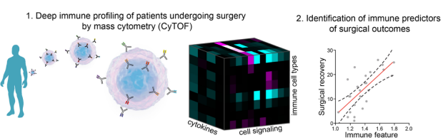
Over the last 5 years, the advent of high dimensional flow cytometry has revolutionized our ability to study and visualize the human immune system. Our group (a collaborative effort with Dr. Nima Aghaeepour and Dr. Martin Angst) combines high parameter mass cytometry (a.k.a Cytometry by Time of Flight Mass Spectrometry, CyTOF), with advanced bio-computational methods to study how the human immune system responds and adapts to acute physiological perturbations. The laboratory currently focuses on two clinical scenarios: surgical trauma and pregnancy.
Deep immune profiling of patients undergoing and recovering from surgery
Using high dimensional mass cytometry, we have recently shown that the signaling behavior of specific innate immune cells measured before surgery in patients’ blood was strongly associated with surgical recovery (Gaudilliere B et al. Science Translational Medicine, 2014; Fragiadakis G et al. Anesthesiology 2015). Prospective validation of reported immune correlates of surgical recovery are underway. Ongoing work in humans and animal models (in collaboration with Dr. Vivianne Tawfik and Dr. David Clark) focuses on the mechanisms by which pre-operative habilitation interventions may alter a patient’s immune state to improve recovery after surgery.
Deep Immune profiling of normal and preterm pregnancy
Our group is an integral component of a multi-disciplinary effort (led by Dr. David Stevenson, Pediatric Department) aimed at understanding the mechanisms of preterm birth, and identifying predictive factors of premature delivery. We have now developed a pipeline and the analytical framework to integrate the single-cell analysis of immune signaling networks by mass cytometry and proteomic profiling of secreted serum factors with the precise phenotyping of pregnancy-related clinical outcomes. In a pilot, cross-sectional study of non-pregnant women, we identified candidate immune signatures that differentiated women with a history of preterm or term pregnancies (Gaudilliere B et al. Cytometry A, 2015). Longitudinal studies in pregnant patients are ongoing to validate these findings.
Dr. Rona Giffard's laboratory studies stroke. Stroke is a devastating problem that is the leading cause of long term neurological disability and the third leading cause of death worldwide. We try to identify ways to reduce ischemic brain injury, and to better understand the interactions between different brain cell types during injury and recovery. Astrocytes and their response to injury is an important focus in the lab. We have found that astrocyte impairment contributes to neuronal injury in global ischemia. Targeting protective strategies to astrocytes leads to markedly increased neuronal survival.
Another area of focus in the lab is the increase in neurogenesis following stroke, and the deleterious effects of inflammation on neurogenesis in this setting. We are studying ways to improve mitochondrial function to increase newborn neuron survival, and modulate inflammation.
MicroRNAs are small noncoding RNAs that reduce translation and thus inhibit gene expression. Recent work shows that microRNAs are regulated in response to stroke, and may play an important role in neuroprotection. We are studying microRNAs that regulate important survival proteins including members of the Bcl-2 family and heat shock proteins. Because each microRNA can target multiple messengerRNAs, this is a way to target several physiologically related genes at once.
Inflammation following stroke while having important necessary roles, can also contribute to worsening injury. We have investigated the importance of IL4 in stroke, and found that it protects male mice from stroke injury. Results in females however, differed. Biological sex differences are seen in differences in the age of stroke and outcome to stroke in patients. We are also investigating sex differences in response to stroke, with a focus on differences in inflammation.
Studies are performed in animal models and primary cultures from brain.
Our laboratory focuses on developing non-narcotic cardiac-safe pain therapeutics and other next generation therapeutics for anesthetic and analgesic care. Further, in order to provide overall better anesthetic care for our patients, we are also examining how common genetic polymorphisms in our patient population we care for may alter anesthetic and analgesic effects of the medications we administer and the post-operative course.
In order to optimize analgesics and limit side-effects, we are also interested in investigating the mechanism of how the nociceptive and cardioprotective signaling pathways are linked. This involves studying the role of nociceptors in cardiac protection and continued interest examining the mechanism of how opioids and volatile anesthetics protect tissue from ischemia-reperfusion injury.
The long-term goal of our research is to provide physiological background information required for the rational design of safer and more effective anesthetics and analgesics.
Hippocampal Research
We investigate the cellular, synaptic and molecular mechanisms of action of central nervous system drugs; especially barbiturates, opiates, anesthetics and other CNS depressants. Electrophysiological recording techniques and selective pharmacological probes are used to investigate the sites and mechanisms of action for CNS depressants. Most of our studies focus on the CA 1 area in rat hippocampal brain slices. Neurons in this brain area are depressed by anesthetics through a combination of pre- and postsynaptic actions on glutamate and GABA mediated neurotransmission.
Theta Research
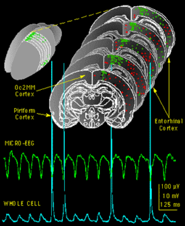
The effects of pharmacological agents on EEG waves generated by the neocortex are also being examined. EEG theta activity (4 to 12 Hz) is one of many rhythms, like alpha and delta (slow wave sleep) rhythms that are altered by anesthetics. Patch clamp and electrophysiological recording techniques are used to look at the effects of anesthetics on carbachol and bicuculline induced theta activity in neocortical brain slices. Anesthetic effects on brain slice micro-EEG activity are correlated to EEG effects seen in animals and humans during anesthesia. Effects on micro-EEG theta activity were shown to involve actions at GABA and glutamate synapses.
Theta activity can be recorded from specific regions (green dots) of cortex in rat brain slices. Comparison of micro-EEG signals and intracellular recordings (whole cell) reveal that the low frequency theta waves (~ 8 Hz) were generated by synchronous synaptic potentials and discharge activity of cortical neurons. The discharge of each cortical neuron appears to contribute ~ 1.0 µV to the micro-EEG signal, so theta activity requires synchronous activity in ~ 100 neurons in each cortical location. Theta activity is known to be important for spacial mapping and may provide a 'binding' mechanism that contributes to the formation of memory in general. When selective populations of neurons are synchronously active they can interact in a Hebbian manner to change the strength of synaptic inputs that are timed at the theta frequency. Theta activity is also known to be particularly sensitive to anesthetic agents at concentrations which block memory formation. Preliminary studies in our laboratory indicate that brain slice theta activity is also depressed by anesthetics and that this depression occurs with a profile similar to in vivo responses.
E-mail Dr. MacIver | http://www.stanford.edu/group/maciverlab/
Please see CAP profile for more details.
Our laboratory investigates the cellular and molecular mechanisms of pain and its control by opioids. We want to identify the neural circuits that underlie pain perception and to resolve the molecular mechanisms by which opioids regulate activity in these circuits to generate analgesia. Our ultimate goal is to uncover new therapeutic strategies to treat chronic pain and opioid-induced side effects. To this end we combine a variety of experimental approaches including molecular and cellular biology, neuroanatomy, electrophysiology, optogenetics and behavior.
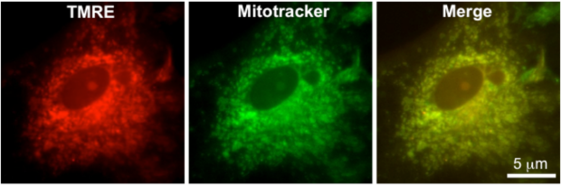
My laboratory is interested in finding new strategies to promote neuronal survival and improve functional outcome following brain injury. The brain consists of several different cell types, the most abundant of which are astrocytes, specialized glial cells that play a vital role in regulating neuronal signaling and homeostasis. All cells depend on mitochondrial function for both normal physiologic functioning and in response to injury; currently we are utilizing microRNAs to simultaneously target multiple pathways that regulate mitochondrial function in both neurons and astrocytes in order to promote cell survival subsequent to noxious stimuli, such as ischemia-reperfusion injury. We employ several in vivo and in vitro techniques, including live cell fluorescent imaging, to assess the cellular response to injury.
My research interests include usage of computational chemistry to develop molecular theories of anesthesia. I collaborate with three groups that use molecular biology to make site-directed mutations in ligand-gated ion channels. Molecular modeling of these channels is used to visualize the effect of mutations and to predict new mutations that will further refine their structure. I also use quantum mechanics calculations to determine the kinds of interactions that are likely to provide binding energy for anesthetic molecules at their sites of action.
Anesthetic Interaction with Membrane Proteins
JIM TRUDELL, PH.D., EDWARD J. BERTACCINI, MD
At the turn of the last century the Meyer-Overton relationship was proposed that relates anesthetic potency to the fat solubility of the anesthetic. This relationship fueled a large academic effort to find the mechanism of anesthetic action. Not surprisingly much attention was focused on determining whether the site of anesthetic action was the lipid layer of the plasma membrane where anesthetics would in some way perturb the lipid bilayer structure. While the results did explain some of the characteristics of anesthetic action there were also many deficiencies. The attention then shifted to the lipid-protein interface where it was thought that anesthetics may disturb the lipid microenvironment around the protein and thereby change the function of the protein. Recently most research has centered on anesthetics directly interacting with membrane proteins and thereby causing a change in their function. Unfortunately anesthetics do not have a high enough affinity for proteins to study the interaction using classical biochemical means. So if the anesthetics were going to play tough and be elusive, the researchers have to be equally inventive. Bring in the big guns: computational chemistry to develop molecular theories of anesthesia.
Drs. Jim Trudell and Ed Bertaccini have been using the methods of bioinformatics, structural biology and computational chemistry to build 3 dimensional models of the various multi-subunit ligand-gated ion channels through which anesthetics are thought to mediate their effects. Much of their work has been focused on the glycine and GABAA receptors found in the brain and spinal cord because they have been shown to play a role in anesthetic action. By doing this extensive modeling study they have developed the first three dimensional visualization of an anesthetic binding site within a clinically relevant protein. Using computational chemistry techniques, they have mapped out this binding site so as to determine the chemical requirements for anesthetic binding. They collaborate with three groups that use molecular biology to make site-directed mutations in these ion channels. The original models are then tweaked to include the modification of the protein sequence and the binding pocket is reexamined. Molecular modeling of these channels is used to visualize the effects of mutations, to predict new mutations to be made and test their properties. This new information will be used to further refine the 3-D structure of the protein.
The results of this work are best seen by looking at the computer images of the receptor model with and without anesthetics. They found a binding pocket in the midst of the protein where there is room for the anesthetic to sit. It seems the anesthetic is held there by weak forces exerted by lipophilic and hydrophilic amino acid residues. If the amino acids within that pocket were modified to make them bigger they could either mimic or block the action of an anesthetic. Even more intriguing-, if the subunits of the receptor are allowed to move in the model, the effect of the anesthetic on the ion channel pore may become apparent.
So will we be better off knowing how inhalational anesthetics work? The answer to that may be yes as knowing how anesthetics work may make it possible to design better anesthetics- ones with more selective and specific actions and ones that might also be more readily reversible.
We are leading the way with two potentially revolutionary approaches to the treatment of chronic pain, namely transplantation and gene therapy. In the first approach, cells taken from the adrenal gland are transplanted on top of the spinal cord through a spinal needle. These cells make and secrete numerous natural analgesic substances, acting like a pump to produce a constant inhibition of pain. Unlike a pump however, these cells are alive, meaning that they keep making and secreting the analgesic chemicals for months.
The primary problems in gene therapy have been the targeting of the right cells, and the duration of the desired effect. We have used a highly modified herpes simplex virus (the kind that causes cold sores) to carry analgesic genes into the pain sensing cells. Because herpes viruses stay in these cells for the life of the host naturally, we should obtain very long lasting analgesic effects using these treatments. Thus, we are making use of the natural proclivity of herpes for entering and staying in the very cells we are interested in. In this way, we target pain treatment to painful areas, and only painful areas. These new approaches to therapy could revolutionize the treatment of chronic pain.
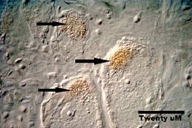
This is a picture of primate dorsal root ganglia neurons demonstrating expression of the opiate analgesic peptide leu-enkephalin after application of a recombinant herpes virus encoding the gene for human preproenkephalin to the skin.