Stanford scientists, using only direct brain stimulation, reproduced both the brain dynamics and the behavioral response of mice taught to discriminate between two different images.
July 18, 2019 - By Bruce Goldman
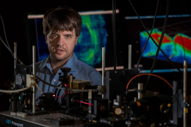
Karl Deisseroth is the senior author of a study describing how he and his colleagues stimulated nerve cells in the visual cortex of mice to induce an illusory image in the animals’ minds.
Steve Fisch
Hallucinations are spooky and, fortunately, fairly rare. But, a new study suggests, the real question isn’t so much why some people occasionally experience them. It’s why all of us aren’t hallucinating all the time.
In the study, Stanford University School of Medicine neuroscientists stimulated nerve cells in the visual cortex of mice to induce illusory images in the animals’ minds. The scientists needed to stimulate a surprisingly small number of nerve cells, or neurons, in order to generate the perception, which caused the mice to behave in a particular way.
“Back in 2012, we had described the ability to control the activity of individually selected neurons in an awake, alert animal,” said Karl Deisseroth, MD, PhD, professor of bioengineering and of psychiatry and behavioral sciences. “Now, for the first time, we’ve been able to advance this capability to control multiple individually specified cells at once, and make an animal perceive something specific that in fact is not really there — and behave accordingly.”
The study, published July 19 in Science, holds implications for obtaining a better understanding of natural information processing in the brain, as well as psychiatric disorders such as schizophrenia, and points to the possibility of designing neural prosthetic devices with single-cell resolution.
Deisseroth is the study’s senior author. Lead authorship is shared by staff scientists James Marshel, PhD, and Sean Quirin, PhD; graduate student Yoon Seok Kim; and postdoctoral scholar Timothy Machado, PhD.
Using optogenetics
Deisseroth, who is a Howard Hughes Medical Institute investigator and holds the D. H. Chen Professorship, pioneered optogenetics, a technology enabling researchers to stimulate particular neurons in freely moving animals with pulses of light, and to observe the resulting effects on the animals’ brain function and behavior.
In the new study, Deisseroth and his colleagues inserted a combination of two genes into large numbers of neurons in the visual cortex of lab mice. One gene encoded a light-sensitive protein that caused the neuron to fire in response to a pulse of laser light of a narrowly defined color — in this case, in the infrared spectrum. The other gene encoded a fluorescent protein that glowed green whenever the neuron was active.
The scientists created cranial windows in the mice by removing a portion of the animals’ skulls to expose part of the visual cortex, which in both mice and humans is responsible for processing information relayed from the retina. The investigators protected this exposed area with a clear glass covering. They could then use a device they developed for the purpose of the study to project holograms — three-dimensional configurations of targeted photons — onto, and into, the visual cortex. These photons would land at precise spots along specific neurons. The researchers could monitor the resulting activity of nearly all individual neurons in two distinct layers of the cerebral cortex spanning about 1 square millimeter and containing on the order of several thousand neurons.
With their heads fixed in a comfortable position, the mice were shown random series of horizontal and vertical bars displayed on a screen. The researchers observed and recorded which neurons in the exposed visual cortex were preferentially activated by one or the other orientation. From these results, the scientists were able to identify dispersed populations of individual neurons that were “tuned” to either the horizontal or vertical visual display.
They were then able to “play back” these recordings in the form of holograms that produced spots of infrared light on just neurons that were responsive to horizontal, or to vertical, bars. The resulting downstream neuronal activity, even at locations relatively far from the stimulated neurons, was quite similar to that observed when the natural stimulus — a black horizontal or vertical bar on a white background — was displayed on the screen.
It’s quite remarkable how few neurons you need to specifically stimulate in an animal to generate a perception.
The scientists trained the mice to lick the end of a nearby tube for water when they saw a vertical bar but not when they saw a horizontal one or saw neither. Over the course of several days, as the animals’ ability to discriminate between horizontal and vertical bars improved, the scientists gradually reduced the black-white contrast to make the task progressively harder. They found that the mice’s performance perked up if the scientists supplemented the visual displays with simultaneous optogenetic stimulation: For example, if an animal’s performance deteriorated as a result of a lowered contrast, the investigators could boost its discrimination powers by stimulating neurons previously identified as preferentially disposed to fire in response to a horizontal or vertical bar.
This boost occurred only when the optogenetic stimulation was consistent with the visual stimulation — for example, a vertical bar display plus stimulation of neurons previously identified as likely to fire in response to vertically oriented bars.
Hallucinating mice
Once the mice had become adept at discriminating between horizontal and vertical bars, the scientists were able to induce tube-licking behavior in the mice simply by projecting the “vertical” holographic program onto the mice’s visual cortex. But the mice wouldn’t lick the tube if the “horizontal” program was projected instead.
“Not only is the animal doing the same thing, but the brain is, too,” Deisseroth said. “So we know we’re either recreating the natural perception or creating something a whole lot like it.”
In their early experiments, the scientists had identified numerous neurons as being tuned to either a horizontal or a vertical orientation, but they hadn’t yet directly stimulated those particular neurons optogenetically. Once the mice were trained, optogenetic stimulation of small numbers of these neurons was enough to get mice to respond with appropriate licking or nonlicking behavior.
The researchers were surprised to find that optogenetically stimulating about 20 neurons — or fewer in some cases — selected only for being responsive to the right orientation could produce the same neuronal activity and animal behavior that displaying the vertical or horizontal bar did.
“It’s quite remarkable how few neurons you need to specifically stimulate in an animal to generate a perception,” Deisseroth said.
“A mouse brain has millions of neurons; a human brain has many billions,” he said. “If just 20 or so can create a perception, then why are we not hallucinating all the time, due to spurious random activity? Our study shows that the mammalian cortex is somehow poised to be responsive to an amazingly low number of cells without causing spurious perceptions in response to noise.”
Deisseroth is a member of Stanford Bio-X and of the Wu Tsai Neurosciences Institute at Stanford.
Stanford’s Office of Technology Licensing has filed a patent application for intellectual property associated with the work.
Other Stanford co-authors of the study are graduate student Brandon Benson; postdoctoral scholars Jonathan Kadmon, PhD, Masatoshi Inoue, PhD, and Hideaki Kato, PhD; life science researcher Cephra Raja; lab managers Adelaida Chibukhchyan and Charu Ramakrishnan; and Surya Ganguli, PhD, assistant professor of applied physics.
The work was funded by the Defense Advanced Research Projects Agency, HHMI, the National Institutes of Health (grants R01MH075957 and P50DA042012), the Simons Foundation, the Wiegers Family Fund, the Nancy and James Grosfeld Foundation, the Sam and Betsy Reeves Fund, the H.L. Snyder Foundation, the Burroughs-Wellcome Foundation, the McKnight Foundation, the James S. McDonnell Foundation and the Swartz Foundation.
Stanford’s departments of Bioengineering and of Psychiatry and Behavioral Sciences also supported the work.
-
 Bruce GoldmanBruce Goldman is a senior science writer in the Office of Communications. Email him at goldmanb@stanford.edu.
About Stanford Medicine
Stanford Medicine is an integrated academic health system comprising the Stanford School of Medicine and adult and pediatric health care delivery systems. Together, they harness the full potential of biomedicine through collaborative research, education and clinical care for patients. For more information, please visit med.stanford.edu.

