Annual Report 2020
> Tele-Ophthalmology
• Advancing optic disc drusen research
• Cross-department team effort
• Vision restoration in glaucoma
• Pursuing excellence through diversity, equity, and inclusion
• Advancing clinical research in the age of COVID-19
• Improving ophthalmologic care through artificial intelligence
• Solving corneal blindness with implantable video technology
Tele-Ophthalmology: Digital care in a digital world
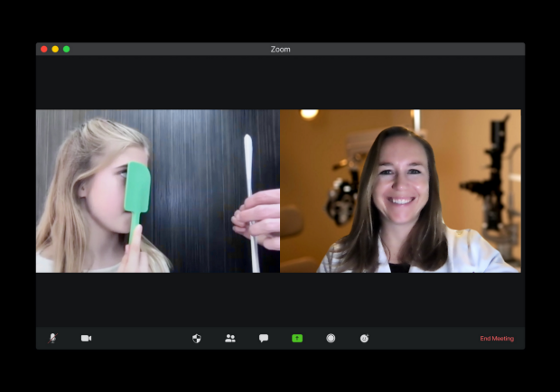
Tawna Roberts, OD, PhD (right), examines a child’s vision during a video visit by using a kitchen spatula to perform a visual acuity test one eye at a time. During video visits, common household items can be used to help perform vision tests.
Traditionally, medical care has been delivered with the provider and patient in the same room at the same time. In-person appointments are vital to conduct certain aspects of the examination, provide in-office treatments, and develop a connection between the provider and patient, even though it can present travel and time burdens on the patient. This year, however, with the coronavirus (COVID-19) public health emergency compelling providers to keep in-person contact to a minimum, alternative solutions have become even more of a pressing need.
In the same way that video conferencing and mobile technologies have revolutionized personal and business communication, their role in health care has also grown significantly. Stanford has long been leading in the utilization of mobile health innovation, but in the face of the COVID-19 pandemic, faculty and staff have had to speed-up this implementation.
To address the ever-growing need for better access to eye care, the Byers Eye Institute at Stanford created a comprehensive offering of “tele-ophthalmology” digital and online resources for patients to meet short-term needs resulting from the pandemic, and also to accelerate into the post-pandemic future. Luckily, the team at Byers Eye Institute had been anticipating this digital future and had already been deploying state-of-the art digital care to infants, children, and adults alike.
Outdoor and mobile testing enable patients to obtain key diagnostic data and then follow up with a video visit with their ophthalmologist. The goal is to provide patients with options that minimize potential exposure to COVID-19 but also provide them with the care they need.
Newborn screening
Darius Moshfeghi, MD, professor of ophthalmology and chief of the retina division at the Horngren Family Vitreoretinal Center, has long been dedicated to preventing and treating blindness in premature infants affected by retinopathy of prematurity (ROP), a terrifying disease in which inappropriate new blood vessels grow in and destroy the retina. The main barrier to successful treatment for ROP is actually inadequate access to qualified screening physicians at neonatal intensive care units (NICUs).
To address this need, Moshfeghi founded the Stanford University Network for Diagnosis of Retinopathy of Prematurity (SUNDROP) program in 2005, and with that also launched Stanford’s Tele-Ophthalmology program, which he directed through 2017. Today, SUNDROP remains the most mature and largest telemedicine program for in-hospital screening of high-risk infants for treatment-warranted ROP in the United States. It has expanded to NICUs at hospitals throughout Northern California, and has spread to two other states, with a dozen or more NICUs in additional states now looking to engage.
SUNDROP has a proven record of reducing blindness and poor visual outcomes from ROP by providing infants in rural and county hospitals with quaternary-level care. At each site, all infants meeting established criteria are screened using RetCam II images taken by hospital staff.
The advantage of this system is that it provides lower cost, high quality care while saving time for the hospital staff, the physicians, and the patient’s family. Moshfeghi has worked with industry partners to develop wide-angle imaging camera technology as well as deep learning classifier platforms for the identification of disease in premature, as well as in healthy term newborn infants. Moshfeghi also serves on the American Academy of Ophthalmology’s Telemedicine Taskforce, which sets practice guidelines for telemedicine for eye care professionals in the U.S.
“The success of the SUNDROP program along with profound changes in imaging and communications technology, regulatory approvals, reimbursement, care guidelines, and patient preferences over the last few years have opened the door to a new paradigm in eye care delivery—one where patient needs can be addressed either remotely, in-person, by a machine, or some combination of these depending on the indication,” Moshfeghi said.
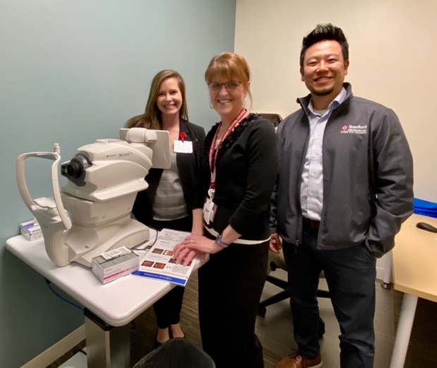
David Myung, MD, PhD (far right), on a visit to one of the remote diabetic eye testing sites in Los Gatos on the day the AI-compatible camera was launched. Also pictured (L to R) are project collaborators from Stanford Health Care and University HealthCare Alliance: Megan Hoehn, Senior Project Manager, Ambulatory Systems; and Maureen Wong, Principal Systems Analyst, IT.
Telemedicine for remote diabetic eye care
In 2017, Moshfeghi passed the role of Director of Tele-ophthalmology to David Myung, MD, PhD, assistant professor of ophthalmology and director of the Ophthalmic Innovation Program. Myung’s goal was to expand the tele-ophthalmology offerings at Byers Eye Institute into adult care, starting with diabetic retinopathy screening. He inherited a set of retinal cameras from Moshfeghi, which had been provided by a donor, and since then has grown what started as a small, one-clinic pilot project into a Bay Area-wide Remote Diabetic Eye Care Network.
The development of this program has been a highly interdisciplinary effort several years in the making through close collaboration between Myung and his retina specialist colleagues at Byers Eye Institute, in particular Theodore Leng, MD, MS, associate professor of ophthalmology and co-director of clinical and translational research. Together, they marshalled key support and contributions from ambulatory care leadership and staff, and information technology and electronic medical records teams, to bring an initial pilot to Stanford’s endocrine clinics. Now, their efforts have accelerated the program’s rollout through Stanford’s primary care and affiliated University Healthcare Alliance community clinics. With expansion of the program to a total of six sites, Vinit Mahajan, MD, PhD, associate professor of ophthalmology and vice chair for research, joined Leng as a remote reader for the captured retinal images through the Stanford Reading Center (STARC).
“It was an extraordinary team effort to set up a network of eye cameras around the Bay Area that provide diabetic patients with the opportunity to have photographs taken of their retinas at their regular primary care or endocrinology appointments, and without the need for dilating drops,” Myung said.
The effort has received accolades and support from both Stanford’s Improvement Capability Development Program led by Diana Do, MD, professor of ophthalmology, and Stanford’s Value-Based Care initiative led by Prithvi Mruthyunjaya, MD, associate professor of ophthalmology.
“If no damage or progression is seen, then patients can continue to follow-up remotely through this program, typically on an annual basis,” Leng said. “Only patients that are found to have evidence of diabetic damage to their retinas are asked to travel to the Byers Eye Institute for further examination and management.”
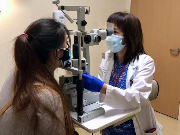
For in-person visits, mandatory masks are worn by physicians and patients. In addition, protective slit lamp shields are used, and surfaces are triple cleaned in between each patient.
Adding artificial intelligence to facilitate tele-ophthalmology
This network of eye cameras now serves as the platform for the newly formed Stanford Tele-ophthalmology Automated Testing and Universal Screening (STATUS) program, established with the goal of enabling patients to more easily check the status of their eye health. As the founding director, Myung is implementing Food and Drug Administration (FDA)-approved artificial intelligence (AI) algorithms and digital health technologies to further expand access to eye care. Now, a fully autonomous AI-based image interpretation algorithm called IDx-DR can read retinal photos and make referral decisions without physician input. Cleared by the FDA, this system is reimbursed by Medicare, and will further boost Stanford’s national quality care metrics for taking care of diabetic patients. These factors set the stage for further expanding the reach of the program by providing exam results and referral decisions to patients immediately while improving workflow and efficiency. Myung is excited about this new paradigm in patient care.
“Leveraging the power of AI for image interpretation provides the option of making routine screenings remote and autonomous, freeing retina specialist colleagues to spend more time with patients who need sight-saving interventions,” Myung said. “Plus, patients get to see their exam results immediately, and can then prioritize the in-person appointments they absolutely should attend while still receiving the care they need.”
The AI-based diabetic eye testing service was launched this year within the Stanford primary care system in Emeryville, to be followed by clinics in Santa Clara and Hoover Pavilion on Stanford campus and at primary care clinics in Los Gatos, Hayward, Oakland, and Pleasanton in early 2021.
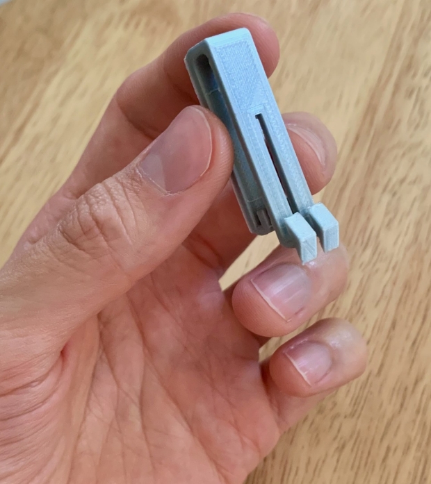
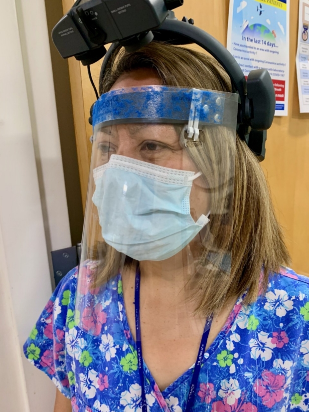
To help with safety precautions during patient visits, resident Jose Davila, MD, designed a small clip that attaches a face shield to an ophthalmoscope to provide extra personal protective equipment during retinal exams.
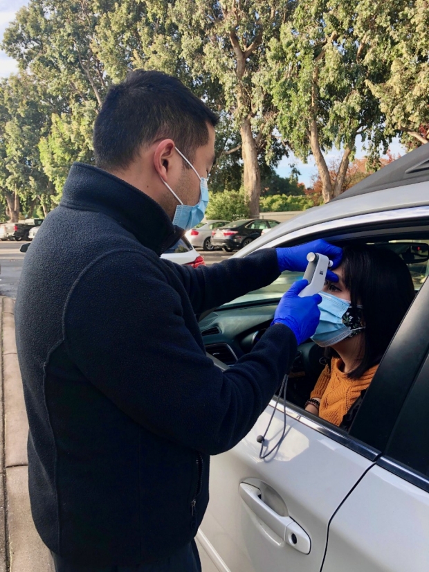
Wen-Shin Lee, MD, established innovative visits wherein patients had a vision check and intraocular pressure measurement from the comfort of their car. Combined with a video visit with their eye doctor, these visits minimized person-person contact while collecting the essential information necessary to allow ongoing management of glaucoma during the pandemic.
Video visits and handheld diagnostics
Prior to COVID-19, Heather Moss, MD, PhD, associate professor of ophthalmology and neurology, saw telemedicine as an ideal platform with which to increase access to care and improve patient experience in her subspecialty of neuro-ophthalmology. Her nascent efforts included opening a satellite eye imaging center in the Stanford Neuroscience Health Center and providing remote image interpretation for neurosurgical and neurological colleagues with specific eye exam questions. During COVID-19, propelled by the need to keep patients and doctors at home while continuing to provide care, Moss led the way in ushering in synchronous tele-ophthalmology appointments at Byers Eye Institute and now directs the Video Visit Program for the department.
Synchronous video visits are accessed via a smartphone application called MyHealth, allowing the patient and provider to see each other. During these video appointments, patients can discuss symptoms, review records sent by their referring provider and ask questions. The physician can perform a limited eye examination including central vision function, peripheral vision, pupils, eye movements, and eye appearance, review in-person testing and discuss management.
Although some subspecialties such as neuro-ophthalmology and oculoplastics are able to heavily utilize video visits (for example, a number of eyelid disorders and neurological disorders impacting vision and eye movements can be evaluated via initial video consultation), other subspecialists need at least some diagnostics that can only be gathered in person. And so, in response to COVID-19, the Byers Eye Institute faculty and staff setup outdoor curbside diagnostic visits.
“Outdoor and mobile testing enable patients to obtain key diagnostic data and then follow up with a video visit with their ophthalmologist,” said Wen-Shin Lee, MD, clinical assistant professor of ophthalmology and a glaucoma specialist. “The goal is to provide patients with options that minimize potential exposure to COVID-19 but also provide them with the care they need.”
In other telemedicine efforts spanning from local nursing homes to villages in rural Nepal, Myung and colleagues have also been able to implement handheld ophthalmic digital health technologies such as smartphone-based cameras and visual acuity tests that further expand access to eye care.
Together, faculty are leading the Byers Eye Institute in what is now arguably the nation’s leading and most comprehensive center for ophthalmologic telemedicine. “The technology we have in the pipeline has so much potential—remote testing, handheld imaging, and AI algorithms— we’re already taking steps into a future that might have been unimaginable even as recently as last year,” Myung said.
What's More?
· Telemedicine process effective at nursing homes: Faculty and researchers from Stanford studied how effective smartphone-based tele-ophthalmology platforms would be for screening patients in the nursing home population. From Stanford, the research group included Myung, Moshfeghi, Mark Blumenkranz, MD, MMS, H.J. Smead Professor of Ophthalmology, Emeritus, Loh-Shan Leung, MD, clinical assistant professor of ophthalmology, Brian Toy, MD, a former resident, and Krystal Lai, a current medical student. Together, they screened patients across 78 Bay Area nursing facilities. They found the smartphone-based platform was effective in detecting multiple eye diseases and could increase eye care access for patients at nursing homes.
· Maternal-Fetal Medicine (MFM) telemedicine for diabetic retinopathy: Malini Veerappan Pasricha, MD, a third-year resident, collaborated with Carolyn Pan, MD, clinical assistant professor of ophthalmology, Myung, and MFM faculty at Santa Clara Valley Medical Center on a year-long study to improve diabetic retinopathy photographic screening for pregnant patients. They studied the telemedicine-based screening model amongst patients in the MFM clinic, increasing the screening rate by 14.7% from the year prior. They found that their model was effective in improving screening rates and providing care in a more cost-effective manner.
· Smartphone-based diabetic retinopathy screening proves more reliable: A group of Stanford doctors ran a diabetic retinopathy study at the Santa Clara Valley Medical Center, comparing mydriatic (pupil dilation) smartphone-based screening versus non-mydriatic (without pharmacologic dilation) table-top camera-based fundus imaging. The group included Myung, Pan, Blumenkranz, Leung, Toy, as well as researchers from other institutions. They found that the smartphone-based screening resulted in both higher quality photos and higher referral rates compared to non-mydriatic photos, making handheld, smartphone-based imaging a promising, cost-effective alternative to conventional methods. A recent study by colleagues in Europe used the same device for diabetic screening in India and came to similar conclusions.
· Could an eye “selfie” help diabetic eye disease patients?: Myung teamed up with Bryan Chiang, a Stanford computer science undergraduate student, to run a clinical trial utilizing an AI algorithm developed by Chiang to non-invasively check glucose through ocular images. This study would help diabetic patients prone to eye disease check their glucose levels without a finger stick or implanted blood glucose monitor. Chiang is grateful that Myung was eager to collaborate with him in the beginning stages of his education. “There is a general energy and enthusiasm toward tackling different health problems that can be felt in the research at Stanford,” Chiang said. “I am thankful to Dr. Myung for creating an interdisciplinary environment that allows me to pursue ideas that can one day help patients.”
· Video visits maintain patient-doctor relationships and increase access to care during COVID-19: Moss and colleagues surveyed the international community of neuro-ophthalmologists and found a rapid adoption of video visits with over 75% utilizing these during the COVID-19 public health emergency. Data quality was the biggest challenge reported by surveyed providers, prompting Moss, along with fellow Byer’s faculty Shannon Beres, MD, and Tawna Roberts, OD, to create training videos to help patients and doctors navigate the eye exam during a video visit.
· Drive-through intraocular pressure visits reduce patient exposure during COVID-19: Lee established innovative visits wherein patients had a vision check and intraocular pressure measurement from the comfort of their car. Combined with a video visit with their eye doctor, these visits minimized person-person contact while collecting the essential information necessary to allow ongoing management of glaucoma during the pandemic.
By KATHRYN SILL
Kathryn Sill is a web and communications specialist for the Byers Eye Institute in the Department of Ophthalmology, at Stanford University School of Medicine. Email her at ksill@stanford.edu.