Annual Report 2021
• An eye-brain connection: Groundbreaking advancements for neurorehabilitation patients
• Shedding light on rare diseases
• Saving vision with gene therapies
• Biorepository: A new key to precision health
> Eye care at all ages: Bringing vision restoration to pediatric patients
• New center tackles rapidly growing myopia prevalence
• My second chance at sight: A patient’s hopeful journey after optic nerve stroke
• Global impact: Generous donors support global health efforts for cataract blindness
• A hopeful view on eyesight: Grateful patient celebrates Dr. Kuldev Singh’s 30th anniversary in 2022
• Fighting blindness across borders
• Stanford Belize Vision Clinic: Training the next generation of eye care providers
• Training for global care: Ophthalmology resident sets up two eye care programs in the Middle East
• Mentorship leads to new gene therapy discoveries
• 3D bioprinting to eliminate corneal blindness
• Big data to transform patient care
Eye care at all ages
Bringing vision restoration to pediatric patients
The Byers Eye Institute at Stanford provides care to patients of all ages, including our pediatric patients from birth to 21 years old, under the auspices of the Lucile Packard Children’s Hospital Stanford (LPCH), with three state-of-the-art facilities. Over the past few years, the pediatric ophthalmology team at Byers has grown to ten faculty committed to preservation and restoration of sight across the full spectrum of eye diseases. For pediatric patients who undergo eye surgery, the journey affects both them and their family.
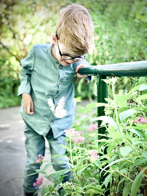
Michael spots a butterfly at the butterfly exhibit.

Michael Reading receives care through the multidisciplinary Cardiovascular Connective Tissue Disorders Program at Stanford Children’s Health and is seen by 21 different specialists.
Super Michael: a living miracle
One inspiring example was recently exemplified in the case of two-year-old Michael Reading, whose surgical team came to talk to him and his parents about his upcoming eye procedure. Michael was prepared, because his parents, Emily and Robert, had already talked through the steps with him. Without skipping a beat, Michael explained to Edward Wood, MD, assistant professor of ophthalmology, that Wood would perform a procedure on his eyes and cover his eyes with bandages as they healed. Then, Michael enthusiastically explained what he was most excited for when his bandages came off: “I’ll see butterflies.”
At a glance, one might not know all Michael and his parents had endured. At birth, Michael was diagnosed with neonatal Marfan syndrome (nMFS), a rare and life-threatening genetic condition weakening all the connective tissue in Michael’s body—most acutely Michael’s heart, lungs, joints and bones, and eyes. By the time Michael was 18 months old, he had already survived three open-heart surgeries. Now, at 26 months old, this eye surgery would be Michael’s fifth major procedure, having received care under 21 different specialists across Stanford.
Every day with Michael is a living miracle. We’ve almost lost Michael three times, but Stanford saved his life. Our family has immense gratitude for the entire Stanford team.
For the eye care team, typical physical manifestations of nMFS include myopia, astigmatism, ectopic lentis, thinning and flattened cornea, glaucoma, retinal detachment, with deeply set and downward slanting eyes. A separate entity from Marfan syndrome, nMFS has much more severe symptoms, with most patients not surviving past the first year.
“Every day with Michael is a living miracle,” Emily said. “We’ve almost lost Michael three times, but Stanford saved his life. Our family has immense gratitude for the entire Stanford team.”
The miracle of Michael’s life and grit has also led to his nickname, “Super Michael,” with the resilience of a superhero.
Michael first became acquainted with the pediatric ophthalmology team at Byers when Deborah Alcorn, MD, professor emeritus of ophthalmology, was brought onto his care team to assess his vision, and after Alcorn’s retirement Scott Lambert, MD, professor of ophthalmology and chief of ophthalmology at LPCH, began overseeing Michael’s vision care. Towards the end of 2020, Michael’s central vision began to deteriorate to the point that his prescription glasses could not compensate. He could only see clearly an inch away from his face and mostly out of his left eye. As his eye’s connective tissue began to weaken, his eye lenses began to take on a sphere-like shape. Because the young brain needs good vision to develop properly, Lambert recommended surgery.

Michael laughing with his parents Emily and Robert in 2020.
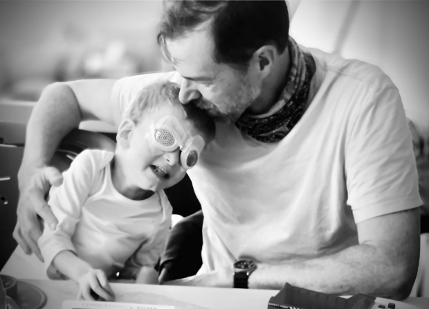
Robert (right) comforts Michael during his recovery period after undergoing bilateral lensectomy and anterior vitrectomy lens surgery and needing to wear protective metal shields.
“Typically, doctors wait until the lenses detach to do surgery on the eyes of nMFS patients,” Lambert said. “In Michael’s case they were still intact, just loose, but with his vision severely worsening, our team felt it important to remove the lenses before the neural pathways in the brain shut down.”
While Lambert had performed many lens removal surgeries, Michael required a specific lens surgery and Lambert asked Wood to perform a bilateral lensectomy and anterior vitrectomy, where both eye lenses and the anterior gel behind the eye pupil are removed. The pediatric ophthalmology team were confident that Michael would see significantly better within a week and be approved for an updated glasses prescription in three months.
After surgery, Michael and his parents remained in the hospital for 24 hours. Metal shields were placed over his eyes for ten days after his surgery, acting as a bandaged barrier to prevent him from touching his eyes as they healed, and his parents also had to administer eye drops every few hours. Soon, Michael began to see again.
Emily still remembers one of the first days after Michael’s eye bandages came off, when he looked at a distant window and spotted a fly. Emily and Robert were in awe, since prior to the surgery he would not have even seen the fly.
“Michael’s ability to see still puts tears in our eyes,” Emily said. “Michael was quickly losing his vision and we didn’t think there were any options, but with the Stanford team and Dr. Wood’s specialized training, Michael has been given the gift of sight. We don’t take this for granted. It’s a miracle to us. Every day we are in awe that Michael can see the world around him.”
Shortly after, they took Michael to a butterfly exhibit, where his prediction that he would see butterflies finally came true.
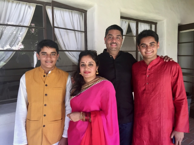
Pictured (L to R): Vignesh, Archana, Mahesh, and Sid.
Stabilizing the cornea, reinforcing hope
Vignesh Sundaram’s hazel eyes are so striking, that throughout his childhood strangers would stop him in public to comment on them. Yet his eyes also told another story, not outwardly visible at the time. Around the age of 12, Vignesh was playing the piano when he began to have difficulty reading the notes on the sheet music.
“To offset my blurring vision, I would push one eye up with my hand until the notes came into focus,” Vignesh said. “Even with the assistance of glasses or soft lenses, my vision was worsening.”
Soon, his mother, Archana Rathnakar, had suspicions that he had keratoconus. Archana was familiar with the initial signs of keratoconus because she had it, as did Vignesh’s older brother, Siddharth (Sid).
The clear front curved surface of the eye, called the cornea, is buttressed by tiny fibers of protein called collagen. A healthy cornea is dome-shaped, but when those fibers break down, the cornea bulges outward into a cone-like shape. Over time, this keratoconus causes the cornea to weaken, leading to vision loss not correctable with glasses. Occurring in about one in every 2,000 persons, the disease typically begins in puberty and if it worsens, a corneal transplant could be needed.
I am thankful for how the team at Stanford not only prevented my vision and cornea from further regression, but that they treated me and my family with great respect, ensuring a smooth process from procedure to follow-up.
Vignesh’s parents took him to the Stanford Health Care - ValleyCare in Pleasanton, where he was referred to Charles Lin, MD, clinical associate professor of ophthalmology. After a series of vision tests, Lin confirmed that Vignesh did have keratoconus, and recommended he receive a newly devised procedure from Edward Manche, MD, professor of ophthalmology and the Director of Cornea and Refractive Surgery at the Stanford Eye Laser Center. Manche specializes in keratoconus treatment and is one of the top performing laser vision correction surgeons in the U.S.
“Vignesh’s cornea had already started thinning from gradual deterioration,” Manche said. “To prevent further thinning that could eventually lead to a future corneal transplant, I recommended a treatment known as corneal cross-linking.”
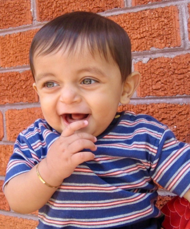
As a child, Vignesh was often stopped by strangers for his striking, hazel eyes.

Vignesh, as a teenager, on vacation in Japan.

Vignesh, now 18, graduated high school in the spring of 2021 from Bellarmine College Preparatory in San Jose.
Cross-linking describes the bonds that hold the collagen fibers together, strengthening the cornea. Manche began by numbing Vignesh’s eye with an eye drop medication, ensuring a pain-free process. Then, Manche used ultraviolet light and riboflavin (vitamin B2), a gel-like fluid, to strengthen the collagen in the cornea. Manche performed the surgery on one eye at a time, the first when Vignesh was 15, and the second a year later.
Following each cross-linking surgery, bandage contact lenses were placed in Vignesh’s eyes for five days, and he was given oral pain medication until the pain subsided. In about ten days the surgery had healed. The surgery successfully stabilized his corneas, with no further deterioration in his vision. Once he was stable, Manche recommended Vignesh see Jill Beyer, OD, clinical assistant professor of ophthalmology to get fit with specialty contact lenses to rehabilitate his vision.
“I learned from Dr. Beyer that correcting vision in a keratoconus patient is extremely different from how you treat vision correction for any other patient,” Vignesh said. “I needed a small contact lens cut in a specific way to meet the needs of my keratoconus.”
Vignesh’s father, Mahesh Sundaram, said that having lived in India, Hong Kong, Australia, China, and now the U.S., their family has had the opportunity to receive patient care in many different countries.
“Stanford is phenomenal in comparison,” Mahesh said. “At Byers, Vignesh received not only eye surgery, but also help through the vision restoration process. The care he received at Stanford by far set the gold standard.”
With both a reinforced cornea and restored vision, Vignesh has returned to doing the activities he loves. Now 18, Vignesh graduated high school this year, where he was a member of the wrestling team and the symphonic jazz band, playing the trumpet and trombone.
“Even while enduring multiple eye complications, Vignesh chose to not view them as a setback,” Archana said. “We are proud of how he continues to be caring towards others and still adventurous towards his goals in life.”
Vignesh has now set his goals on a college degree. In the fall of 2021, he began his fall semester at Pepperdine University, majoring in psychology, confident that with his vision issues addressed he can continue to excel in the classroom and beyond.
“I am thankful for how the team at Stanford not only prevented my vision and cornea from further regression, but that they treated me and my family with great respect, ensuring a smooth process from procedure to follow-up,” Vignesh said.
By KATHRYN SILL
Kathryn Sill is a web and communications specialist for the Byers Eye Institute in the Department of Ophthalmology, at Stanford University School of Medicine. Email her at ksill@stanford.edu.