Annual Report 2021
> An eye-brain connection: Groundbreaking advancements for neurorehabilitation patients
• Shedding light on rare diseases
• Saving vision with gene therapies
• Biorepository: A new key to precision health
• Eye care at all ages: Bringing vision restoration to pediatric patients
• New center tackles rapidly growing myopia prevalence
• My second chance at sight: A patient’s hopeful journey after optic nerve stroke
• Global impact: Generous donors support global health efforts for cataract blindness
• A hopeful view on eyesight: Grateful patient celebrates Dr. Kuldev Singh’s 30th anniversary in 2022
• Fighting blindness across borders
• Stanford Belize Vision Clinic: Training the next generation of eye care providers
• Training for global care: Ophthalmology resident sets up two eye care programs in the Middle East
• Mentorship leads to new gene therapy discoveries
• 3D bioprinting to eliminate corneal blindness
• Big data to transform patient care
An eye-brain connection
Groundbreaking advancements for neurorehabilitation patients
Department faculty use augmented reality computer simulation to heal the visual pathway.
Our vision depends not just on our eyes, but on the full visual pathway from eye to brain. Injury to this visual pathway at any point leads to vision loss, and oftentimes to loss of independence. Eager to discover therapeutics and diagnostic methodologies for patients suffering from these injuries, faculty at the Mary M. and Sash A. Spencer Center for Vision Research at the Byers Eye Institute at Stanford are leading the way in innovative research through departmental and multidisciplinary collaborations. Clinician-scientists have recently discovered a myriad of findings relating to this eye-brain connection with implications that may change the way we diagnose and treat concussion-related vision disorders, strokes of the optic nerve or brain (including non-arteritic anterior ischemic optic neuropathy [NAION]), visuo-motor dysfunction, and other diseases, translating their discoveries directly into clinical care to optimize patient outcomes and overall quality of life. (To read about a patient’s experience with NAION, see “My second chance at sight: A patient’s hopeful journey after optic nerve stroke”, page 18).
Joel Alan Imventarza, MD, a postdoctoral research scholar, collaborates with Joyce Liao, MD, PhD, and tests out an eye movement recording monitor.
Vision restoration in patients with visual pathway injuries and abnormalities
The brain needs feedback from the eyes to move our extraocular muscles, which keep the two eyes aligned on their target. Misalignment of the eyes, called strabismus, leads to debilitating symptoms including double vision, loss of depth perception, and in children, failure of the visual centers in the brain to develop properly. Focusing on how this eye-brain connection can be leveraged towards vision rehabilitation efforts in pediatric patients, Tawna Roberts, OD, PhD, assistant professor of ophthalmology, is involved in an American Academy of Optometry-funded U.S. multi-center clinical trial on vision therapy for intermittent exotropia (IXT). This effort is led by Angela Chen, OD, MS, FAAO, associate professor at the Southern California College of Optometry at Marshall B. Ketchum University. IXT occurs when one eye turns outward, causing misalignment of the eyes and loss of depth perception when the eye is deviated. It is the most common form of childhood-onset exotropia. If left untreated, it can have a severe negative impact on a child’s quality of life.
The group’s study is the first clinical trial to assess the efficacy of vision therapy incorporating objective eye movement recordings and biofeedback. Surbhi Bansal, OD, clinical assistant professor of ophthalmology, is serving as the vision therapist for Stanford’s patients on this study and Jen Haensel, MS, PhD, a postdoctoral research fellow in Roberts’ Lab, is the lead for the objective eye movement recordings. This study will allow the team to directly test how much vision therapy helps each patient.
“This is a great opportunity to objectively study eye alignment and accommodation simultaneously in a clinical disease where eye alignment is the primary outcome measure,” Haensel said.
Advanced eye movement recordings measure each child’s eyes, capturing both eye position and accommodation at a high sampling rate, to objectively assess the efficacy of the treatment. The goal of the vision therapy is to provide patients with the awareness of when their eyes are misaligned and to train them to re-align their eyes using biofeedback.
“This form of therapy allows patients to progress at a pace that is comfortable for them,” Roberts said. “Our vision therapist will monitor them throughout the process. The program also builds in difficulty as patients improve their skill level. Our hope is that this therapy will produce long-term, lasting results.”
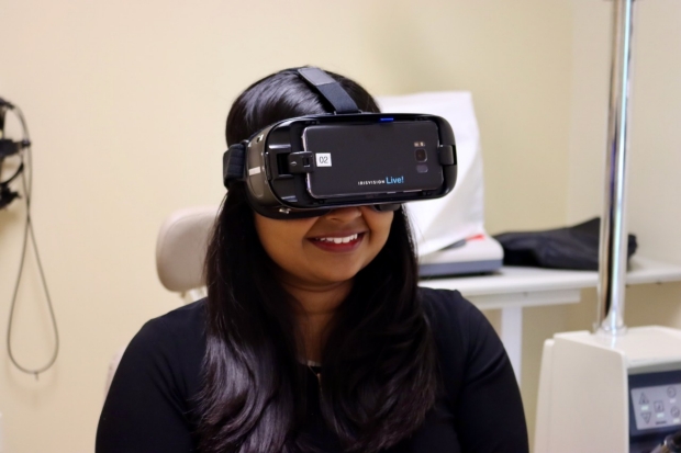
Jeffrey Goldberg, MD, PhD, the Blumenkranz Smead professor and chair of ophthalmology; Joyce Liao, MD, PhD, professor of ophthalmology and neurology; and Roberts are also involved with work being done to heal the visual pathway through augmented reality (AR). AR uses computer-simulation to generate a real-world environment through three-dimensional images, sensory stimuli, and sound. Unlike virtual reality (VR), which requires a headset to immerse users in their simulation, AR can be used on a camera or smartphone or with special transparent “smart glasses”, and add to a user’s visual experience, rather than fully substitute for the surrounding world.
Moving forward, our goal is to develop effective ways to enhance visual function in patients through pharmacology including medications and eye drops, as well as nonpharmacologic ways including augmented reality, virtual reality, and visual rehabilitation exercises.
These computer-simulated devices are now making their way into the clinic with hopes of addressing different vision impairments and diseases. While still in the pilot stage, they are manipulating what a patient sees through AR smart glasses, that look like large sunglasses.
“These are essentially smart glasses that are adjustable,” Liao said. “If you’re reading or looking at a digital device, we could either enhance the image by altering the brightness or increasing the contrast in a specific part of the visual field, or shift the image in front of one eye.”
This would also allow them to align the eyes for a patient with double vision by adjusting an image to exactly what the patient needs to see. This differs from traditional glasses, which require lenses to be set at a specific prescription and is not instantaneously adjustable, whereas AR allows for flexibility in prescription strength. In addition, AR could potentially be used to train patients with double vision through vision stimulation exercises.
“Moving forward, our goal is to develop effective ways to enhance visual function in patients through pharmacology including medications and eye drops, as well as nonpharmacologic ways including augmented reality, virtual reality, and visual rehabilitation exercises,” Liao said. “Both methods are promising.”

Joyce Liao, MD, PhD, and Tawna Roberts, OD, PhD, serve as the co-directors of the Stanford Human Ocular Motor Lab. Pictured (L to R) are lab faculty: Jeffrey Goldberg, MD, PhD; Surbhi Bansal, OD; Liao; Roberts; and Khizer Khaderi, MD, MPH. The lab also includes other research team members and students.
Foveation and visuo-motor dysfunction in brain diseases
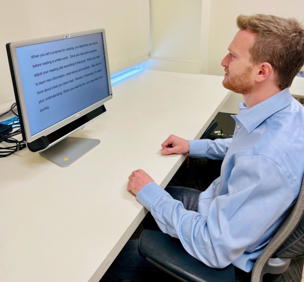
Another way the eye and brain must work together is on foveation—the process by which we make rapid eye movements to align an object of interest on our fovea, the most visually sensitive part of the retina in the center of our vision. Foveation allows us to find a person’s face in a crowd or look at a bird as it passes by. The process of repeated eye movement and foveation allows the eyes to take rapid snap shots of the world like a camera, and this information is sent to the brain for interpretation. The Stanford Human Ocular Motor Lab uses high-speed video infrared eye trackers to capture such visuo-motor patterns to quantify and understand how patients move their eyes during normal activities like reading or visual search.
“Understanding how this visuo-motor axis may cause dysfunction should allow for better design of treatments and new opportunities for visual rehabilitation,” Liao said.
In a new streamlined process, patients can now be seen in clinic and taken to the eye movement recording lab in the same day. An eye movement test displays a computer screen with an eye tracker placed at the bottom of the screen that monitors the patient’s eyes by having them look at different words and numbers. This provides a non-invasive way to capture quantitative data in these patients. The eye movement information is then compared to other patient data, including demographics, neuro-ophthalmology assessments, traumatic brain injury severity, and neuropsychology surveys.
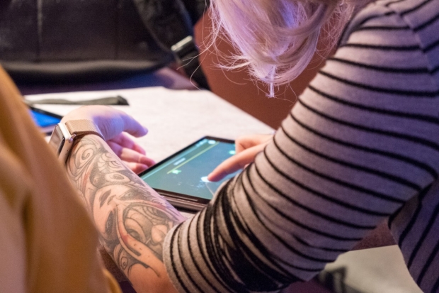
Khizer Khaderi, MD, MPH, and members of the Stanford Human Perception Lab and the Vision Performance Center, attended the Riot Games Pro Esport All-Star Event in Las Vegas, Nevada in December 2020. Khaderi uses video games embedded with algorithms to optimize vision.
In one exciting new direction, Liao, Roberts and Bansal are using eye movement recordings to discover treatment options for patients with Parkinson’s disease, a brain disorder that leads to bodily tremors and other movement deficits. Patients with Parkinson’s disease tend to have abnormal eye movement patterns. They have measured eye movement behavior in hundreds of subjects with a variety of vision and eye movement issues. Their research is now some of the first to investigate how eye and brain diseases affect visual function, such as reading or watching scenery on a digital screen.
“This breakthrough is critical for eye movement exams, which were historically one of the more challenging parts of the clinical eye exam, so hopefully this new process will make diagnosis, monitoring, and designing appropriate treatment for patients easier,” Liao said.
Going forward, Liao said their goal is to develop an automated, artificial intelligence (AI) platform that can diagnose eye movement disorders. This would open the door for not only helping those with Parkinson’s disease, but those with other neurodegenerative diseases and brain injuries as well.
This future goal might be met by Khizer Khaderi, MD, MPH, clinical associate professor of ophthalmology and a neuro-ophthalmic surgeon. Prior to joining Stanford, Khaderi started his first technology company, iSportGames, using video games embedded with algorithms to stimulate cells in the retina. The result was improving sport performance by 22% in athletes by optimizing their vision. His second startup, Vizzario, utilizes a computerized AI platform to develop new methods of measuring visual function via the connection between the eye-brain-body.
Khaderi’s idea stems from wanting to improve the traditional eye exam with advancements in technology. One of these methods, the Vision Performance Index (VPI) is being utilized to monitor and predict visual, cognitive, and motor performance for professional athletes (traditional & Esport), patients with both ocular and neurodegenerative disease, and workplace productivity in technology companies. VPI is also powering the success of an international AI performance lenswear company, Monokül.
Now at Stanford, Khaderi has founded the Stanford Human Perception Lab and the Vision Performance Center, where they are building and applying technologies to help patients optimize their eye-brain-body performance through rehab and training.
“Sensory technologies can improve the visual health for a variety of patients,” Khaderi said. “I want to see innovative technology not only aid in the healing process for those with eye injuries, but to also enhance vision, so that patients can have their best potential vision long term.”
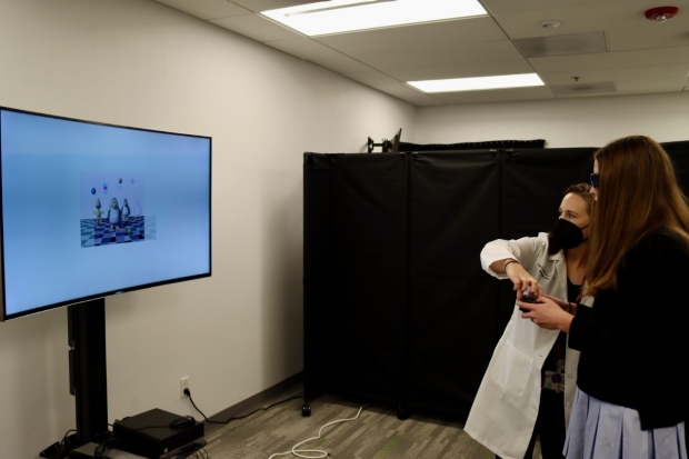
Pictured (L to R): Tawna Roberts, OD, PhD, helps Georgia Hutchinson perform a vision rehabilitation exercise to address her traumatic brain injury.
Healing concussion-related vision disorders in adolescents and adults
When Georgia Hutchinson was 13 years old, she took an elbow to the head during a water polo game. Georgia, who had always excelled in academics, noticed something off at school the next day. She called her mother, Christine Hutchinson saying her memory was fuzzy and she felt sick to her stomach. Christine took Georgia to her pediatrician who diagnosed her with a mild traumatic brain injury (mTBI), more commonly referred to as a concussion, an injury that effects approximately 3.6 million people in the U.S. every year. When Georgia’s symptoms did not heal on their own after a few weeks, her pediatrician referred her to the Stanford Pediatric Concussion Clinic.
By this point Georgia’s vision had begun to suffer. Vision problems are common after mTBI with many patients reporting blurred and/or double vision, ocular pain, or difficulty focusing on close items. For children, mTBI is particularly problematic because their brains are not yet fully developed and their vision symptoms often impact their ability to read and do schoolwork, even furthering the negative effect of the injury.
Georgia was seen by Roberts and Gerald Grant, MD, FACS, Botha Chan Endowed professor. Grant serves as the Stanford Division Chief of Pediatric Neurosurgery and the Director of the Pediatric Concussion Clinic. They monitored Georgia, having her perform at-home exercises to help her better focus and align her eyes, and within six months her normal visual function was restored.
Georgia, now 16, has traded water polo for a new sport of rowing and is considering pursuing a future career in neuroscience or biomedical engineering.
“Georgia was always interested in science and the medical field, and her interactions with Dr. Grant and Dr. Roberts only strengthened her interests,” Christine said. “Both doctors provided the utmost care for Georgia. I continue to recommend friends’ children to the concussion clinic because of the excellent professionalism and empathy we were shown at Stanford.”
On the research side, Roberts and Grant are leading a team of clinicians and researchers across multiple subspecialties at Stanford to better understand vision disorders and their related symptoms in adolescents with mTBI. Liao, Bansal, Beres, and Heather Moss, MD, PhD, associate professor of ophthalmology and neurology, and other Stanford faculty in different departments are also involved with the study, which is funded by the National Eye Institute.
“A critical barrier in optimizing the management of patients with concussions is that we don’t know how long it takes for vision symptoms to resolve after the traumatic brain injury, which has an impact on medical decision making for when to refer patients for vision care,” Grant said. “By the time it is decided that the symptoms are not getting better, and the patient is referred for a vision examination, many months may have gone by.”
To tackle this problem, the study will take place at eight clinical centers across the United States and one in Canada. Each clinical center is made up of a team of doctors who manage the mTBI and a team of doctors who manage the vision problems related to the mTBI. While COVID-19 has pushed back the project’s start date, the team began patient recruitment in fall 2021.
In addition to studying pediatric mTBI, Roberts and Bansal are involved in research funded by the United States Department of Defense looking at mTBI in Stanford University varsity athletes in hopes to better understand vision disorders in our servicemen and women who incur injuries while on the battlefield. When these vision problems don’t heal on their own, vision rehabilitation may be needed.
“We’re using high quality camera systems to measure eye focusing and eye movements, as well as EEG to measure the brain’s response to visual motion. These study paradigms allow us to see if we can manipulate various visual inputs to the brain to better understand how the eyes move and focus on those inputs and ultimately how the injured brain processes the visual inputs,” Roberts said.
These research findings will then be transferred into the clinical setting through a collaboration with the Stanford Brain Performance Center (SBPC) directed by Jamshid Ghajar, MD, PhD, FACS, clinical professor of neurosurgery and Angela Lumba-Brown, MD, clinical associate professor of emergency medicine. SBPC is located in the Lacob Family Sports Medicine Center located at the Arrillaga Center for Sports and Recreation. Geoff Abrams, MD, assistant professor of orthopedic surgery, has a team of Stanford therapists and physicians at the Lacob Family Sports Medicine Center in varying specialties who tailor the treatment given to each mTBI athlete. Bansal, who performs vision evaluations and provides treatment for vision disorders associated with mTBI in patients from the Stanford Neuroscience Health Center, said she is excited to expand and enhance clinical care to the Stanford Varsity athletes who will benefit from this research.
“Eye movements, tracking a moving target or fixating on a target when the person is moving, produce headache, dizziness, nausea and fogginess, which are the main mTBI symptoms,” Ghajar said. “Roberts’ groundbreaking research will shed light on the mechanism in mTBI eye movements that are producing impairments in brain function so that we can better treat patients. We are excited to work together as a multidisciplinary eye-brain team to better diagnose and treat mTBI patients.”
By KATHRYN SILL
Kathryn Sill is a web and communications specialist for the Byers Eye Institute in the Department of Ophthalmology, at Stanford University School of Medicine. Email her at ksill@stanford.edu.