Annual Report 2021
• An eye-brain connection: Groundbreaking advancements for neurorehabilitation patients
• Shedding light on rare diseases
• Saving vision with gene therapies
> Biorepository: A new key to precision health
• Eye care at all ages: Bringing vision restoration to pediatric patients
• New center tackles rapidly growing myopia prevalence
• My second chance at sight: A patient’s hopeful journey after optic nerve stroke
• Global impact: Generous donors support global health efforts for cataract blindness
• A hopeful view on eyesight: Grateful patient celebrates Dr. Kuldev Singh’s 30th anniversary in 2022
• Fighting blindness across borders
• Stanford Belize Vision Clinic: Training the next generation of eye care providers
• Training for global care: Ophthalmology resident sets up two eye care programs in the Middle East
• Mentorship leads to new gene therapy discoveries
• 3D bioprinting to eliminate corneal blindness
• Big data to transform patient care
Biorepository
A new key to precision health
The lack of laboratory models for human eye diseases is a roadblock to translational research, but it drove Vinit Mahajan, MD, PhD, associate professor of ophthalmology and vice chair for research, to forge a scientific path that promises to lead to medical discoveries. The Byers Eye Institute Biorepository has been established since 2018, and allows the collection of thousands of biospecimen samples, creating a repository of data never before assembled.
By studying the fluid from human eyes undergoing surgery, Mahajan and co-director Prithvi Mruthyunjaya, MD, MHS,associate professor of ophthalmology, are leading a department-wide collaboration to support personalized medicine breakthroughs with an immediate impact on patient care. Faculty are unveiling disease mechanisms, finding drugs to repurpose into effective new treatments, and developing novel small molecule therapies.
Mahajan and Mruthyunjaya sat down to share their experiences.
Q: What prompted you to create the Biorepository?
VM: We knew eye tissues and fluids discarded during surgery hold the molecular clues researchers need to cure eye diseases. Researchers could benefit from using these “liquid biopsies” to identify key protein biomarkers and answer critical clinical questions: What’s the right drug for a patient? Is this an infection or autoimmune condition? Is it hereditary? Will this eye cancer metastasize? Will a patient go blind?
PM: Protein biomarkers can take the guesswork out of patient care and potentially guide clinical therapy and clinical trials. Analyzing the molecular makeup of diseased tissues can explain drug therapy failure and reveal rare disease mechanisms in humans.
Q: What impact has the Biorepository had on the department’s research?
VM: A major goal is to expand and support the application of translational proteomics, the large-scale study of proteins, throughout the department. We are also collecting stem cells and DNA for genetic testing.
PM: Using diseased tissue from patients, Mahajan and I were the first to identify proteins from inside the eye that predict survival risk in patients with ocular melanoma, a lethal eye cancer. For the first time we will be able to tell which patients are at highest risk and need the most aggressive monitoring and treatment. Our findings also point to personalized patient therapies and could aid disease surveillance.
VM: My lab identified a new metabolic therapy approach to treat genetic eye disease and macular degeneration. Dr. Andrea Kossler is studying thyroid eye disease and ocular surface inflammatory diseases; Drs. Robert Chang, Wen-Shin Lee, Yang Sun, and Jeffrey Goldberg are looking at glaucoma; Dr. Joyce Liao is studying optic nerve disease; Dr. Charles Lin is investigating corneal infections; Dr. Yang Sun is also using human stem cells collected from patients with rare diseases; and partnerships with industry are helping to focus their drug development programs on the right targets.
THE PROCESS
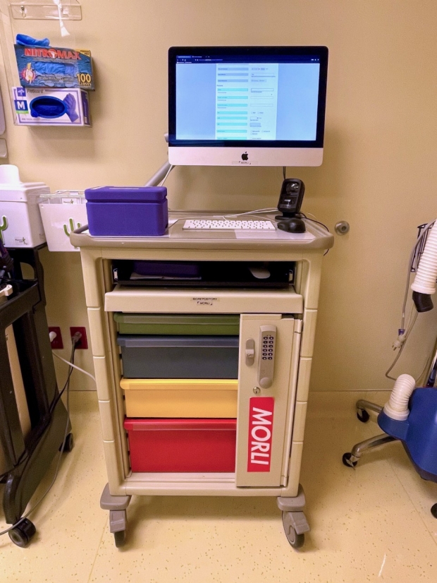
STEP 1: Ophthalmic surgical specimens are processed and preserved on MORLI, a laboratory on wheels computer system with a bar code scanner.

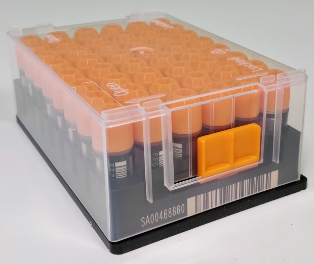
STEP 2: Flash freezing and transporting specimens to an -80 degree Fahrenheit laboratory freezer allows for long term storage and preservation.

STEP 3: Research findings can then be transferred to the clinical setting.

Q: What are some of the obstacles to preserving tissues for molecular research?
VM: Linking the operating room and research labs was a major challenge that we solved. It requires collaborations between OR staff, surgeons and research scientists, clinical research coordinators, protein biochemists, and bioinformaticians. The Mobile Operating Room Laboratory Interface (MORLI) now links the operating room with the laboratory, bringing people together who would normally never cross paths.
Q: Tell me more about MORLI.
VM: The MORLI is a “laboratory on wheels” designed with all the required instruments and devices. This makes it possible for the surgical team to immediately process specimens in the operating room and maintain specimen integrity. The MORLI has a computer, bar code scanner, and all the lab supplies necessary for processing and preserving tissue samples.
PM: Collecting and tracking ophthalmic surgical specimens with the MORLI has facilitated the collection of high-quality research samples whose molecular profiles are well-preserved. So far, we have collected over 3,000 human surgical samples. We now perform advanced molecular analyses on samples straight out of the operating room.
Q: What happens once the specimens are collected?
VM: Typically, specimens are flash frozen and transported to a -80 degrees Fahrenheit laboratory freezer for preservation. Flash freezing tissue allows us to save and study the molecules that would otherwise degenerate in minutes. In some instances, we’ve actually done the biochemical tests just outside the operating room door.
Q: How are specimens tracked?
VM: Surgical staff use iPads to obtain consent from patients using patient privacy protection measures. Key clinical data is entered into a database linked to the barcoded sample tubes and samples are stored in secure freezers.
Q: What broader impact does the Biorepository have on research?
PM: We have a system for creating an efficient biorepository of human eye tissue samples, which can be applied to any type of biological tissue. These samples are shared with researchers at Stanford and collaborators across the country and around the world.
VM: A collection of human tissue samples so carefully collected and preserved is priceless. Converting our research findings into clinical diagnostics and therapies and expanding this platform into multi-center studies will further our department’s commitment to providing patient care based on the broader goals of precision health.
By MARYANN MAHAJAN
MaryAnn joined Mahajan's lab in 2008. She earned her B.A. in English at the University of California, Berkeley and attended graduate school at the University of California, Los Angeles where she earned a secondary teaching credential. She is a writer and editor and also performs histological phenotyping of mouse eyes.