Annual Report 2021
• An eye-brain connection: Groundbreaking advancements for neurorehabilitation patients
• Shedding light on rare diseases
• Saving vision with gene therapies
• Biorepository: A new key to precision health
• Eye care at all ages: Bringing vision restoration to pediatric patients
• New center tackles rapidly growing myopia prevalence
• My second chance at sight: A patient’s hopeful journey after optic nerve stroke
• Global impact: Generous donors support global health efforts for cataract blindness
• A hopeful view on eyesight: Grateful patient celebrates Dr. Kuldev Singh’s 30th anniversary in 2022
• Fighting blindness across borders
• Stanford Belize Vision Clinic: Training the next generation of eye care providers
• Training for global care: Ophthalmology resident sets up two eye care programs in the Middle East
• Mentorship leads to new gene therapy discoveries
> 3D bioprinting to eliminate corneal blindness
• Big data to transform patient care
3D bioprinting to eliminate corneal blindness
The cornea is the clear, outermost part of the eye. Like a camera lens, it focuses light onto the retina which acts as a sensor for the images projected onto it. As a living tissue without blood vessels, the cornea can heal from minor injuries, but major injury or disease leads to scarring that blocks light, blurs images, and can lead to blindness.
Over 12 million people worldwide suffer from corneal blindness. Corneal transplants can restore sight, but a shortage of tissue donors accompanied by the economic and in some cases cultural barriers to harvesting, screening, and delivery, means that less than 2% of patients globally have access to this surgery.
Bioprinting allows us to greatly scale up what is currently a one-to-one donor-to-recipient procedure and potentially generate a supply of engineered tissue needed to meet the world’s demand for transplantable corneas.
“For the majority of the millions of people worldwide who are blind due to corneal scarring, only a fraction of these patients have a chance at seeing again because of the lack of accessible donor corneal tissue,” Myung said.
To develop alternative and more scalable treatments to replace traditional corneal transplant, David Myung, MD, PhD,assistant professor of ophthalmology and, by courtesy, chemical engineering, and director of the Ophthalmic Innovation Program, and his lab are collaborating with colleagues in the School of Engineering to create a biosynthetic alternative to donor tissues.
In this interdisciplinary collaboration with Sarah Heilshorn, MS, PhD, professor of materials science and engineering, and their co-mentored chemical engineering PhD students Sarah Hull and Lucia Brunel, they are using a process known as 3D bioprinting to fabricate and grow cornea tissue in the lab. In bioprinting, a bioink is dispensed from a nozzle to deposit cells within a biological matrix having a geometry specified by computer-aided design (CAD). The team is printing engineered corneal tissue of the same dimensions of human donor corneas, which has about the same material volume as a drop of water.
Their unique bioink technology, which they’ve called UNIversal Orthogonal Network (UNION) bioinks, has been further engineered to encapsulate and print corneal stem cells in the lab using a modified 3D printer.
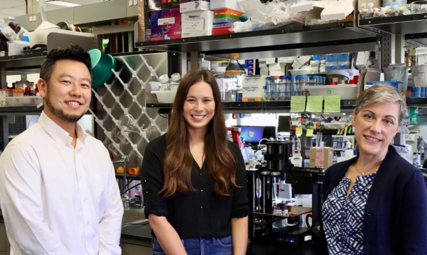
Pictured (L to R): David Myung, MD, PhD; Sarah Hull; and Sarah Heilshorn, MS, PhD, are using 3D bioprinting technology to create transplantable engineered corneas to substitute for cadaveric donor corneal tissue, which is in short supply worldwide.
Realizing the platform’s potential, the team is also using it to pursue an on-demand approach to corneal repair, where the bioink is applied directly to a patient’s wounded eye to fill and heal a corneal defect. Myung likens this approach to biological spackling paste used to fill defects and holes in drywall, but with regenerative effects. Using a handheld dispenser that they have designed, a single droplet of their bioink can fill a deep stromal defect of the cornea as a viscous liquid. That liquid then solidifies into a soft gel, reconstructing the smooth, transparent outer contour of the cornea, and allowing a protective epithelial layer to grow completely over its surface.
“Bioprinting allows us to greatly scale up what is currently a one-to-one donor-to-recipient procedure and potentially generate a supply of engineered tissue needed to meet the world’s demand for transplantable corneas,” Myung said.
The UNION bioink platform has been engineered so that it does not require toxic chemicals, catalysts, light, or heat that many other polymer crosslinking technologies require and also be applied without sutures. They have published on the platform recently in top scientific journals including Biomaterials, Scientific Reports, and Advanced Functional Materials. Myung is also collaborating with Jennifer Cochran, PhD, Shriram professor and chair of bioengineering, to incorporate growth factors that can help accelerate healing and reduce scarring.
“The challenge is to not only engineer a material that starts off like transparent corneal tissue, but also stays clear as it is remodeled and replaced.” Myung said. “If fibrosis occurs during the healing process, then a scar will form, and you have to start all over.”
Myung’s interest in biomaterials for the cornea began during his MD/PhD years when he was awarded one of the first Interdisciplinary Bio-X Fellowships at Stanford. As new crosslinking modalities emerged during the course of his ophthalmology residency training, he realized that the technology to enable sutureless corneal repair was now readily available and just had to be harnessed in the right way.
To support their research, Myung and colleagues have obtained funding from the Bio-X interdisciplinary scholars program to support Hull’s PhD work, as well as major grants from the National Eye Institute. Moving forward, Myung hopes to garner additional funding to more rapidly translate his findings quickly into the clinic.
By KATHRYN SILL
Kathryn Sill is a web and communications specialist for the Byers Eye Institute in the Department of Ophthalmology, at Stanford University School of Medicine. Email her at ksill@stanford.edu.