Invented at Stanford
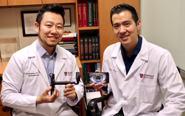
David Myung, MD, PhD, (left) and Robert Chang, MD, display the Paxos Scope, an ophthalmic camera system for smartphones they helped invent.
The Stanford Department of Ophthalmology has a rich history of innovation and academic accomplishment dating back more than 100 years. In both engineering and biological sciences, Stanford has often led the translation of lab-based advances into clinical diagnostics and therapeutics.
“Stanford is well known for spinning out technologies and inventions and for collaborating with partners in Silicon Valley,” said Mark Blumenkranz, MD, H.J. Smead professor emeritus of ophthalmology.
Blumenkranz, chair of ophthalmology from 1997 to 2015 and the inaugural director of the Byers Eye Institute from 2010 to 2015, has been a driving force behind a number of successful innovations, adding to a list of Stanford contributions to ophthalmology that started in the 1960s. In the late 1960s and early 1970s, two volunteer faculty members, Hunter L. Little, MD, and H. Christian Zweng, MD, helped invent the first slit lamp-based ophthalmic laser delivery systems. In the mid-to-late 1990s, novel research in retina and cornea moved from Stanford labs to patient care. Starting in 1998, multiple cutting-edge diagnostic, therapeutic and surgical tools were developed in the laboratory of Daniel Palanker, PhD, professor of ophthalmology and director of the Hansen Experimental Physics Laboratory, and then commercialized in conjunction with pharmaceutical and device companies in Silicon Valley, helping millions of patients worldwide. (See timeline on pages 8-9.)
Many of these “invented at Stanford” successes have leveraged the breadth and depth of the Stanford community, such as the invention and subsequent commercialization of TrueTear, a neurostimulation device that activates tearing from the lacrimal gland and is now clinically available for patients with dry eye disease. Michael Ackermann, PhD, a Biodesign fellow (see below) and then vision research postdoctoral fellow with Palanker, led a development team in conjunction with others including Andrea Kossler, MD, assistant professor of ophthalmology, and Christopher Ta, MD, professor of ophthalmology. In addition to prototyping and validating the technology’s efficacy clinically, they formed a company Oculeve that was later acquired by Allergan.
“Being a biodesign fellow and then vision research fellow and postdoctoral fellow at Stanford was an incredible and pivotal two years for me,” Ackermann said. “That time helped me make the transition from scientist to entrepreneur, all while gaining ophthalmology experience under the training of phenomenal faculty.”
Blumenkranz, who helped commercialize Oculeve, agrees.
“Innovation is critical to advancing progress, because the world is moving at a rapid pace in terms of information technology, molecular biology, imaging, and globalization,” Blumenkranz said. “Leveraging those advances allows us to deliver care that results in better access and outcomes for patients.”
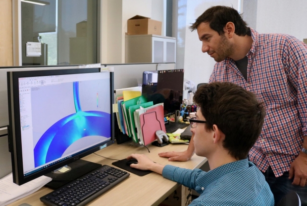
Frank Brodie, MD, MBA, and David Buickians work together on understanding the behavior of a novel intraocular implant using a computer-aided design simulation.
“Innovation is critical to advancing progress, because the world is moving at a rapid pace in terms of information technology, molecular biology, imaging, and globalization. Leveraging those advances allows us to deliver care that results in better access and outcomes for patients.
Establishing the Ophthalmic Innovation Program
Recognizing a successful formula linking identification of unmet needs to discovery, invention, and commercialization, Blumenkranz established the Ophthalmic Innovation Program in 2015. This educational program provides a unique, one-year post-graduate fellowship for clinicians and scientists. Fellows are taught the necessary steps to take an idea from conception to clinical use. David Myung, MD, PhD, assistant professor of ophthalmology and, by courtesy, of chemical engineering, has co-directed the program since its inception.
“Each year the fellow participates in hands-on projects, formal coursework, close mentorship, and networking and internship opportunities with members of the department, other Stanford departments, Silicon Valley innovators, and colleagues at the Center for Devices and Radiological Health at the Food and Drug Administration (FDA),” Myung said.
This year’s fellow is Frank Brodie, MD, MBA, whose research includes development of a novel intraocular implant to assist in cataract surgery and custom-printed 3D glasses to help the vision of children with craniofacial abnormalities develop correctly. Through a non-profit organization he started, The Loving Eyes Foundation, these glasses are then given to the children at no cost.
“This fellowship is unlike any other,” Brodie said. “It is an incredible, unprecedented opportunity to do translational research, learn the process of innovation hand-in-hand with regulatory science, and develop transformative ophthalmic products.”
Synergies with Biodesign
Several ophthalmology faculty members are alumni of the Biodesign Faculty Fellowship (BFF) program within the Stanford Byers Center for Biodesign. Robert Chang, MD, assistant professor of ophthalmology, was part of the second BFF class from 2015–2016 and continues to teach medical innovation focused around digital health. One of his original contributions is the creation of the health design sprint, a week-long project-based learning experience leading students through the challenges in starting a health care company.
“We are not just training excellent doctors and researchers but also passing on the innovation method and the entrepreneurial spirit to the next generation of leaders in ophthalmology and vision research,” Chang said.
ADDITIONAL DEVICES
We couldn't fit a number of devices in this year's annual report. For those interested, here is more information on devices that have been invented within or in collaboration with the Ophthalmology Department.
Pulsed Electron Avalanche Knife
Daniel Palanker, PhD, professor of ophthalmology and director of the Hansen Experimental Physics Laboratory, and his lab team developed an extremely precise electrosurgical instrument, the Pulsed Electron Avalanche Knife (PEAK), with the theoretical capacity to cut at the most minute levels – a single cell while avoiding the usual collateral damage caused by conventional electrosurgical instruments like the Bovie, the prior standard of care. Confinement of the exposed part of the electrode to a few micrometers in width and pulse duration to a few microseconds limited the interaction zone to the size of a single cell. On the other hand, the large length of the blade provided convenience of a macroscopic surgical instrument, similar to a scalpel. Collateral damage zone of this “Plasma Blade” did not exceed a single cell width. Having the capacity to adjust the pulse duration on the fly to milliseconds enabled deeper heating - sufficient for hemostasis. This instrument was successfully tested in retinal and cataract surgeries, and later commercialized, with primary applications in the fields of plastic surgery, ENT, orthopedic, spine surgery, and other fields. The device is now manufactured and distributed by Medtronic (Plasma Blade).
- o 1998- Invented by Palanker
- o 2005- Commercialized by a startup PEAK Surgical, Inc., formed by Blumenkranz and Palanker
- o 2011- Acquired by Medtronic
Laser Systems
PASCAL Laser System
Blumenkranz and Palanker, in collaboration with residents and fellows they trained, created the PASCAL (PAtterned SCAnning Laser) for retinal therapy, which has since been adopted by the industry as the preferred method of precise retinal photocoagulation. Retinal laser photocoagulation used to be a painful and time-consuming procedure, and because of this was often broken up over multiple sessions lasting one to two hours or more in the aggregate for patients with more advanced cases of diabetic retinopathy. The PASCAL laser dramatically shortened this procedure to a single 5-15 minute visit, with much less discomfort for the patient in the vast majority of patients. In addition, this system helped to minimize or in some cases even eliminate the deleterious side effects of conventional photocoagulation and greatly increase clinical efficacy.
Femtosecond laser system for cataract surgery
Until recently, cataract surgery was performed manually, which limited precision of the intraocular lenses’ centration and its overlap with the anterior capsule and was prone to difficulties in cases of poor visibility, weak zonules in elderly patients, and very elastic capsule in pediatric cases. This optical coherence tomography-guided femtosecond laser system is capable of all the cutting of the cornea, lens capsule, and segmentation of the lens itself. After capturing the 3D image of the eye, the system defines placement of all the cutting patterns, which reduces the dependence on surgical skills, tissue properties and visibility and also allows for heretofore unforeseen levels of precision in cutting. It also reduces the ultrasonic energy during lens emulsification, thereby providing for reduced damage to corneal endothelial cells. The system (CATALYS) is now manufactured by Johnson & Johnson and is in clinical practice worldwide. Their patent and the excellent clinical results paved the way to introduction of several similar systems by other major manufacturers (Alcon and Bausch & Lomb).
- o 2004- A Silicon Valley startup OptiMedica Corporation licenses the PAttern SCAnning Laser System (PASCAL) invented by Blumenkranz and Palanker at Stanford to dramatically improve the precision, speed and comfort of retinal photocoagulation.
- o 2004-2005-The CATALYS Femtosecond Laser System (CATALYS Precision Laser System) for cataract and refractive surgery also invented by Palanker and Blumenkranz in conjunction with physicists and engineers at OptiMedica is patented, prototyped, and subsequently, commercialized by OptiMedica
- o 2010 – The PASCAL retinal laser photocoagulator is acquired by Topcon Medical Systems, Inc.
- o 2013- Abbott Medical Optics acquires OptiMedica and the CATALYS Femtosecond Laser System
- o 2017- Johnson & Johnson acquires Abbott Medical Optics
Adverum Biotechnologies
In 2005, a graduate student Tom Chalberg began conducting research on gene therapy using pulsed electroporation in Palanker’s lab. Together with Blumenkranz, they developed a concept of biofactory, which is a transfection of the patient’s own tissue in order to secrete a protein for suppression of the vascular endothelial growth factor signaling to treat macular generation. They then founded a company called Avalanche Biotechnologies. This company went public in 2014 (NASDAQ).
Later Palanker and Chalberg developed a laser-based approach to down-regulate protein expression, which led to greater precision and fine tuning of gene therapy affects. Blumenkranz served as the co-founder and chairman of the board until October 2016. In 2016, the company changed its name to Adverum Biotechnologies.
- o 2005- Chalberg and Palanker begin conducting research on gene therapy using pulsed electroporation
- o 2006- Blumenkranz, Palanker, and Chalberg develop and patent the concept of a retinal biofactory using new gene therapy techniques for in vivo production of synthetic proteins and monoclonal antibodies which is the genesis of a new startup company Avalanche Biotechnologies
- o 2013 – Palanker and Chalberg conduct research on modulation of the transgene expression in retinal pigment epithelium (RPE) using lasers, and this patent is licensed to Avalanche Biotechnologies
- o 2014 – Avalanche Biotechnologies goes public on NASDAQ
- o 2016 - Avalanche Biotechnologies acquires additional gene therapy programs and changes name to Adverum Biotechnologies
PRIMA – photovoltaic retinal implant
Retinal degenerative diseases can lead to blindness due to loss of photoreceptors, while the inner retinal neurons are relatively well-preserved. Electrical stimulation of the inner retinal neurons allows reintroducing information into the visual system, thereby enabling restoration of sight. Palanker developed a high-resolution photovoltaic retinal prosthetic system, in which processed images from a video camera are displayed on video goggles and projected through the eye optics onto the retina using pulsed near-infrared light. Each pixel in the subretinal photovoltaic array converts light into electric current to stimulate the nearby neurons. High irradiance near-infrared reflectance light does not affect remaining photoreceptors and thus allows full utilization of the residual peripheral vision, and is also of sufficient energy to be sufficient to create an electrical signal in adjacent neurons, something that was not possible with older subretinal passive systems that relied upon ambient illumination. Optical projection of the images into the eye preserves the natural link between eye movements and visual information and allows scaling up the number of electrodes to thousands. Lack of any wiring greatly simplifies the surgery and allows implanting multiple modules via small retinotomy to tile a large visual field. A clinical trial with this system (PRIMA) developed in collaboration with Pixium Vision, confirmed the feasibility of significant improvements in patients who had lost most of their central vision from retinal degeneration, and demonstrated the highest acuity of prosthetic vision achieved in humans so far - 20/460. Now the Palanker lab is working on reducing the pixel size down to 20 um, which may support visual acuity up to 20/80 in theory. If successful, this would make photovoltaic implants applicable to millions of people who lost central vision due to age-related macular degeneration and for whom there are currently no alternative approved therapies.
- o 2005- Palanker lab invents a new type of photovoltaic retinal prosthesis
- o 2013- The technology is licensed to Pixium Vision
- o 2014 – Pixium Vision goes public (Euronext Paris)
- o 2018 – First proof of concept and safety clinical trial with PRIMA implant in age-related macular degeneration (AMD) patients is initiated in Europe and the United States
TrueTear
Perhaps one of the greatest successes that best summarizes the synergy between the department and Stanford Biodesign is the invention of TrueTear, as mentioned previously. The invention was developed during the first year the department interacted with biodesign and was geared towards patients with dry eye disease.
Michael Ackermann, PhD, a past biodesign fellow and then vision research fellow and postdoctoral candidate in Daniel Palanker’s, PhD, professor of ophthalmology and director of the Hansen Experimental Physics Laboratory, lab, led a development of electrical stimulation of the lacrimal gland, as well as the afferent nerves in the lacrimal system. They also worked in conjunction with others from the department, including Andrea Kossler, MD, assistant professor of ophthalmology, and Christopher Ta, MD, professor of ophthalmology. Ackermann’s group demonstrated that their approach resulted in a dramatic increase in tear production. Together with Blumenkranz, they formed a company Oculeve, which developed a neural stimulator, TrueTear, and demonstrated excellent results of this technology in clinical tests. For the first time, not only the volume of tear fluid has increased, but also the objective indicators of the corneal surface health and subjective assessment of the symptoms were greatly improved. The company was later acquired by Allergan, and the system, TrueTear, was approved for clinical use world-wide.
Not only did this interaction give rise to a successful invention, but it leveraged future opportunities for interactions between the department and biodesign. This device continues to be utilized in the clinical setting and showcases what advancements can be made when strong interactions happen.
“Being a biodesign fellow and then vision research fellow and postdoctoral fellow at Stanford was an incredible and pivotal two years for me,” Ackermann said. “That time helped me make the transition from scientist to entrepreneur, all while gaining ophthalmology experience under the training of phenomenal faculty members.”
- o 2010- The concept of neuromodulation to stimulate abnormally low production of tears in dry eye patients is developed and patented by Biodesign students spearheaded by Michael Ackermann working in conjunction with members of the ophthalmology faculty.
- o 2011– Prototype designed, built, and evaluated in Palanker lab during time Ackermann is a postdoctoral fellow in the department of ophthalmology.
- o 2011- A start-up company based on this neuromodulation approach, Oculeve is formed and licenses the intellectual property from Stanford OTL.
- o 2015- Oculeve acquired by Allergan and the technology named TrueTear
- o 2017 Oculeve’s TrueTear is approved for human use by the FDA
SightBook
- o 2010 - Blumenkranz and Palanker develop a smartphone-based app for evaluation of visual function at home, and web-based connection of patients with physicians.
- o The app (SightBook) is commercialized by DigiSight Technologies – a company formed by Blumenkranz and Palanker to help advance the nascent field of ophthalmic mobile health.
Paxos Scope
When Myung began his ophthalmology residency at Stanford in 2012, he noticed the camera quality on smartphones was improving tremendously. He began working with Robert Chang, MD, assistant professor of ophthalmology, to create a small device that could take medical grade images of the eye. Together they co-invented the EyeGo system, an ophthalmic camera system for smartphones that captured both anterior and posterior images, which was the predecessor device for what is now known as Paxos Scope.
After teaming up with a former graduate mechanical engineering student, Alexandre Jais, who printed 3D models of their invention, they added a built-in light source into the device to help with seeing the back of eye. They then began collaborating with DigiSight Technologies (now Verana Health) who helped register the device with the Food and Drug Administration (FDA) and bring it into the clinic.
“This device helped build the foundation for some of the processes we implement in the Ophthalmic Innovation Program, including interactions with the FDA and other steps that are necessary to translate a new technology into the clinic,” Myung said.
“When we developed the Paxos Scope, there already were some portable devices on the market that coupled smartphones to existing, expensive ophthalmoscopes,” Myung said. “Our invention simplified the optics and reduced cost by coupling phones directly to a single lens. It is the first and only FDA-registered smartphone-based device designed to take pictures of both the front and the back of eye.”
The Stanford Byers Center for Biodesign awarded a Spectrum MedTech seed grant to fund the initial work, and later the invention was licensed to DigiSight Technologies who ultimately commercialized the device in the US.
Paxos Scope has been used globally through Chang’s studies in India and Myung’s collaborations with the Himalayan Cataract Project and the Tilganga Institute of Ophthalmology in rural Nepal, where the device is being studied as a way to screen patients for eye diseases in remote village settings. By providing high quality imaging in low resource settings and the ability for remote evaluation by ophthalmologists, the Paxos Scope has demonstrated its potential to dramatically improve care for patients around the world.
- o 2012- Myung and Chang begin brainstorming ophthalmic camera system for smartphones
- o 2013-2014- Myung, Chang, Blumenkranz and Alexandre Jais patent and publish on Paxos Scope for both anterior segment and retinal photography
- o 2014- Licensed to DigiSight Technologies (now Verana Health) and further developed to improve access to eye for patients in underserved areas and in out of the office locations including the home, workplace and other hospital departments including the emergency room and primary care settings for assistance in the evaluation of patients with trauma, diabetes, and other acute emergencies.
- o 2015- Paxos Scope registered with the FDA via 510(k) pathway as an ophthalmic camera system
AVA, Inc.
- o 2016- Chang’s invention created with faculty in the Biodesign Faculty Fellowship Program, Eric Sokol, MD, associate professor of obstetrics and gynecology and, by courtesy, urology, and Jan Liphardt, associate professor of bioengineering
By KATHRYN SILL
Kathryn Sill is a web and communications specialist for the Byers Eye Institute in the Department of Ophthalmology, at Stanford University School of Medicine. Email her at ksill@stanford.edu.