Jump to Story
- 2023 Letter From the Chair
- The Paths to Clinical Care
- Precisely Yours
- Improving Vision
- Drug Discovery
- Meet Dr. M.E. Hartnett
- Game On
- Al Revolution
- Life, Uninterrupted
- Sight Restored
- Giving Mission
- Modern Day Textbook
- Training the Next Generation
- Select Awards and Honors
- Why Give
- Residents and Fellows
Precisely Yours
As precision medicine grows, patients benefit most
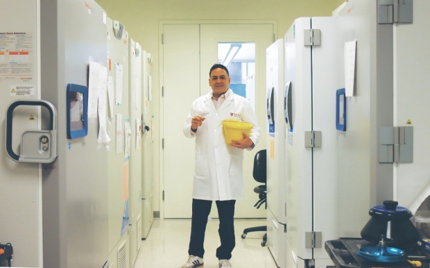
Dr. Vinit Mahajan co-created the Byers Eye Institute's biorepository.
PRECISION HEALTH is the latest buzzword in medicine, promising to help doctors better tailor care to the individual and allow people to proactively address potential health issues before they become a problem. But it’s not always clear what that looks like in practice.
At the Byers Eye Institute at Stanford, precision health is more than just a buzzword. Many of the precision health initiatives at Byers hold the promise of preventing—or even curing—diseases of the eye, staving off or reversing vision loss for patients. The innovative programs range from new data collection procedures that make diagnosing disease faster and more accurate to developing revolutionary imaging technologies and expanding access.
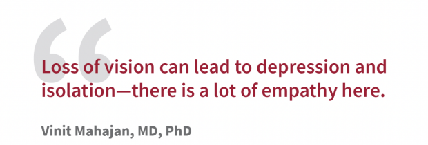
When Vinit Mahajan, MD, PhD, professor of ophthalmology, talks about precision health, you can practically feel the excitement radiating from him. Many patients are perfectly healthy except for their vision problems, he says, but “vision is our primary way of engaging with the world around us. Loss of vision can lead to depression and isolation—there is a lot of empathy here.”
Diving into Data
A key pillar of the Mary M. and Sash A. Spencer Center for Vision Research is the Byers Eye Institute Biorepository, which Mahajan helped create and now co-directs. This collection includes blood samples, cheek swabs, liquid from tears, and tissues and fluids taken from patients’ eyes during surgery. Once in the biorepository, research teams can sequence DNA and measure several thousand proteins to mine for data overlooked in the past.
“With eye fluid samples, we can get a very precise picture of which cells inside the eye are being affected,” Mahajan said. “These are fluids that would otherwise be thrown away, but by analyzing them, we can diagnose diseases much better in our patients. We also learn which molecules we should design therapies for.”
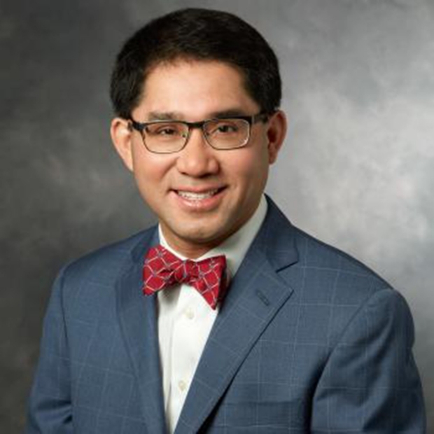
Quan Dong Nguyen, MD, MSc
Careful review of a patient’s medical history, combined with aids like the data from the biorepository, can narrow the search for appropriate treatments, according to Quan Dong Nguyen, MD, MSc, professor of ophthalmology, and by courtesy, of medicine and pediatrics. Nguyen’s biorepository studies have proven invaluable for the diseases he manages.
“Now that we can detect the proteins that our medicines are targeting, we can detect disease and individualize treatments,” he said. “This is a major avenue to advance both research and patient care.”
Nguyen, who specializes in uveitis and ocular inflammatory diseases as well as retinal disorders, also looks for signs of diseases that could lead to eye inflammation. “We are now better able to detect and define local eye disease and systemic rheumatic diseases, caring for the whole patient, not just their eyes,” he said.
Precision health can also reduce cultural bias in health care, Nguyen added. Tailoring tests to specific patients across races and ethnicities can offer important information about predispositions and inflammatory markers that might otherwise be overlooked.
Enhancing the image
Meanwhile, Alfredo Dubra, PhD, professor of ophthalmology, is developing imaging techniques that provide sharper images of the eye in a way that goes far beyond other current imaging technology.
Dubra is focused on early diagnosis and treatment, which are especially important in eye diseases because most cells in the retina can’t reproduce; once they’re gone, they’re gone for good.
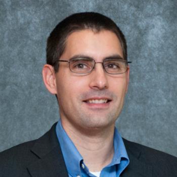
Alfredo Dubra, PhD
“Current ophthalmoscopes provide views of wide retinal areas," Dubra said. "This approach is quick, but not very sensitive,” Dubra said. “Many cells could die before any changes can be detected. We are trying to develop novel instrumentation to image the retina as if we were looking through a microscope, seeing individual cells, and even their movement.”
More precise measurements could reveal pathological changes over weeks instead of months to years, allowing physicians to adjust treatments faster.
Another problem Dubra’s lab is targeting is how to capture retinal images in people with nystagmus, an extreme form of involuntary eye movement where the eyes flicker back and forth. “By developing new eye tracking and movement correction technology in real-time, we can capture retinal images and improve care for these patients who are not benefiting from any form of today’s retinal imaging,” he said.
Indeed, early diagnosis is the key to most effectively treating or preventing disease in the first place. “The goal of precision health is to detect patients at risk and keep them from developing vision loss in the first place,” says Jeffrey Goldberg, MD, PhD, Blumenkranz Smead professor and chair of ophthalmology at Stanford.
"One advantage we have is that the eye is right out there in front, and we can leverage high-resolution imaging and novel technologies to actually see very fine cell structures, even cell metabolism, and use these as biomarkers of health and disease," he said.
BY BARBARA EGBERT
Barbara is a freelance writer for the Byers Eye Institute at Stanford.