Community Profiles
In Community Profiles we highlight the activities and accomplishments of our medical and bioscience education community members. We hope this content will help others identify common interests across departments and specialties, learn about the vast array of educational activities taking place across Stanford Medicine, and lift up the valuable contributions of our educators. Contact Alicia DiGiammarino at adigiamm@stanford.edu if you have content to feature in a future Community Profiles segment.
Dr. Reena Thomas
Clinical Associate Professor, Department of Neurology and Neurological Sciences
Reena Thomas earned her PhD in Molecular Immunology before entering medical school, always knowing she wanted to be a physician scientist. As a Clinical Associate Professor in Neuro Oncology at Stanford Medicine, she has accumulated over a decade of experience serving as PI of several clinical trials and research programs. In addition to her scholarly efforts, Dr. Thomas has been committed to supporting diversity and inclusion in medicine, and has served in a number of leadership roles that have allowed her to have a positive impact in this area across the school. This fall, Dr. Thomas will step into the role of Senior Associate Dean for Medical Education in the School of Medicine. This appointment is a next step for Dr. Thomas who is currently the Associate Dean for the Office of Diversity in Medical Education, as well as the Vice Chair for Diversity and Inclusion in her department.
ROLES
Adult Neuro Oncology Fellowship Program Director (2014-2021)
Education Chief of Neuro Oncology, Department of Neurology (2017-2020)
Director of Diversity and Inclusion, Department of Neurology (2017-2021)
MSTP and PSTP Berg Scholars Admissions Committees
Associate Dean, Diversity in Medical Education, schoolwide (2020-Present)
Vice Chair, Diversity and Inclusion, Department of Neurology (2021-Present)
ADVICE
Be of service
Dr. Thomas was aware that she had many mentors, teachers, and sponsors who helped to shepherd her through her education and into her career, and she has always been oriented toward giving back, committed to helping others coming after her as she herself had been supported. Early in her career, motivated by her committement to be of service, Dr. Thomas identified informal opportunities to do so—mentorship of trainees from diverse backgrounds, outreach around recruitment. Through these informal, volunteer experiences, she built her networks, her understanding of the medical education landscape at Stanford, and her experience, preparing her to move into roles in which she could be of service in a more formalized way. She explains that by looking for where she could be of service, she was able to build a path that ultimately lead to bigger, more impactful roles in areas that were most meaningful to her.
Build your skillset
Dr. Thomas also described joining committees earlier in her career that would allow her to develop skills or knowledge she felt she would need later in her career. For example, by becoming involved in the MSTP admissions committee, Dr. Thomas gained a deep understanding of what goes into decision-making for MD-PhD student admissions. Then, as her skills and knowledge evolved, she was ready to take roles with greater reach such as Faculty Director in the Office of Diversity in Medical Education (ODME), which ultimately led to stepping into the role of Associate Dean for this office. As Associate Dean, Dr. Thomas expanded ODME’s scope to include support for residents and fellow trainees in addition to working with UME and pre-college students, creating a continuum of pre-faculty career development.
Say ‘yes’ to what you love (and be OK with saying ‘no” sometimes too)
The example of Dr. Thomas’ evolving role with ODME highlights one final bit of advice she offers: say ‘yes’ to the things you love and follow what interests you most. From the beginning, Dr. Thomas has said yes to opportunities that allowed her to mentor and support others while simultaneously learning the ins and outs of medical education. When asked how she decides when to say ‘yes’ versus ‘no,’ Dr. Thomas says to remember that the career in medical education is a marathon, not a sprint, and with this in mind, she does encourage people to remember that it is OK to say ‘no.’ And how to know when to say ‘yes’? Look for activities and opportunities that align with what matters most to you. In addition to helping you build your professional networks and opening up new opportunities, this approach ultimately allows you to build a career path that is meaningful and rewarding for you.
Dr. Rebecca Ivancie
2023 - 2024 TMA Innovation Grant Awardee
Teaching Non-Clinical Skills Essential to a Career in Community Hospital Medicine: A Curriculum for Skills Beyond the Bedside
Pediatric Hospital Medicine (PHM) Fellowship aims to develop clinician-leaders who assure the best care of children in both university-based and community hospitals. PHM Fellowship includes a minimum 4-week community hospital medicine (CPHM) rotation. However, little is known about the strengths and weaknesses of current training in preparing fellows for work in community settings. An informal needs assessment with leaders from the AAP SOHM Fellowship Directors Task Force and Community PHM Subcommittee highlighted potential training gaps for PHM fellows, specifically in non-clinical skills (NCS) essential to careers specifically in CPHM. PHM Fellowship graduates who currently work in CPHM may offer unique insights regarding knowledge and skill gaps. Our study’s objective is to explore perspectives of graduated PHM fellows working in CPHM regarding what they learned and what gaps they see in learning non-clinical skills in community hospital settings during fellowship.
We conducted a qualitative study using semi-structured virtual focus groups of PHM fellowship graduates working in CPHM. Focus group transcripts were open coded independently by five researchers who then met to iteratively create and refine a codebook and identify content domains using consensus-driven methods. We conducted five focus groups with fourteen participants total.
Twelve domains emerged around training gaps, with nine for non-clinical skills and three for clinical skills. Non-clinical topics specifically geared toward CPHM that participants felt were inadequately covered in fellowship yet important for community practice included: Advocacy, Autonomous Practice with Limited Resources, Business of Medicine, Career Planning and Advancement, Education and Scholarship in CPHM, Health Systems Practice, Interdisciplinary Team Dynamics and Communication, Leadership and Administration, and Perception and Value of CPHM (Table 1). While our study focused on non-clinical skills, three domains also emerged regarding clinical skill gaps: Neonatal medicine, Emergency and Critical Care Skills, and Triage and Transfers.
Based on our needs assessment, there are important educational opportunities in PHM fellowship to further highlight and expose fellows to some clinical and many non-clinical aspects of CPHM practice during their training. Future directions include curricular development and mentoring programs to best prepare fellows for CPHM careers.
Click here to read more about Dr. Ivancie's TMA Innovation Grant and about our other Grantees!
Dr. David Hartmann
2023 - 2024 TMA Innovation Grant Awardee
Engineering ChatGPT Prompts to Build a Case-Based Learning Library in Neurology
Surveys of Stanford Neurology residents show that we prefer to learn by working in teams to discuss real cases, an approach called team problem-based learning (PBL). Just as in real patients, in our education we enjoy working as a team to review a patient’s history, exam, and clinical data (such as MRI or other imaging) to arrive at a diagnosis and treatment plan. Unfortunately, team problem-based learning exercises are difficult and time-consuming to create, which leads to the overwhelming majority of our didactics being delivered as powerpoint lectures.
In my project sponsored by the Teaching and Mentoring Academy, I am validating the use of ChatGPT as a tool to convert published patient cases into team problem-based learning exercises. As many readers know, ChatGPT is a publicly-available large language model that uses machine learning to create text that varies based on input “prompts.” So far I have learned that ChatGPT does a stellar job of creating team problem-based learning exercises, and next I will survey neurology residents in order to find out which ChatGPT prompt structure produces the most accurate and educational PBL exercises.
Click here to read more about Dr. Hartmann's TMA Innovation Grant and about our other Grantees!
Dr. Susan Marie Lang
2023 - 2024 TMA Innovation Grant Awardee
Establishing a high-fidelity laparoscopic training curriculum for trainees in Obstetrics & Gynecology
In our study, we are enhancing the laparoscopic surgical proficiency and confidence among Obstetrics & Gynecology (Ob/Gyn) resident trainees at Stanford University Hospital. Using a combination of a newly created curriculum, expert content videos, and a high-fidelity porcine simulation model, the project aims to assess the impact on skill enhancement and perceived confidence through validated evaluation metrics. By conducting pre- and post-training assessments, the effectiveness of the simulation model in improving laparoscopic skills is being evaluated. Furthermore, the project extends its focus beyond residents to include more advanced procedures among surgical fellows, thereby fostering a culture of continuous learning and skill development within the institution.
By establishing a high-fidelity simulation model and curriculum as a standard training tool within the Ob/Gyn department, the project not only addresses immediate training needs but also lays the groundwork for sustained improvement in surgical education in women’s health. Anticipated outcomes include the establishment of a robust training resource with high translational potential, contributing to improved patient outcomes and fostering collaborative efforts within the medical community. Overall, this project aims to not only enhance surgical skills but also cultivate a culture of excellence and innovation in medical education.
Click here to read more about Dr. Lang's TMA Innovation Grant and about our other Grantees!
Dr. Zihan Zhou
Postdoctoral Education Fellow
Development and application of a rapid robust 3D-MRF with fast online recon in a longitudinal pediatric sample
Recent advances in novel quantitative MR Fingerprinting (MRF) have been developed by our lab to allow for rapid high-resolution quantitative T1 and T2 mapping. However, the clinical application of MRF faces challenges: (A) The lengthy image process (reconstruction) time hampers real-time viewing of results, impeding image quality checks and prompt head-motion monitoring to avoid patient recall. (B) Field-inhomogeneities (B0/B1) degrade the images, necessitating additional calibration scans that prolong scan and reconstruction.
In our study, we upgraded the MRF sequences and reconstruction algorithm. This not only reduces the reconstruction time (to within 4-minutes), but also enhances the result robustness to B0/B1 inhomogeneity and head-motion. The proposed acquisition/reconstruction pipeline has been validated on over 100-scans performed across longitudinal neuroscientific settings on pediatric samples with plan for open-source distribution soon. After validating its repeatability and reproducibility on a longitudinal pediatric study, in the next phase of our study we plan to analyze the MRF data which was performed on each subject before and after an intense reading-intervention to address a neuroscientific research question: can quantitative T1 and T2 measurements be used to study intervention-induced brain development?
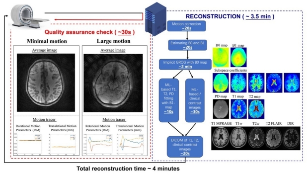
Figure 1. Fast online reconstruction pipeline. The left panel shows the quick assurance check which is displayed online in 30s after the scan completion. Minimal and large motion cases are shown, where a re-scan would recommend for large motion cases, while for smaller motions, the build-in motion-aware reconstruction is capable of providing high quality reconstruction. The reconstruction pipeline generates T1, T2, PD as well as synthetic contrast images robust to B0- and B1-inhomogeneity and motion artifacts. The total pipeline takes 4 minutes.
Dr. Albaraa A. Basfar
Postdoctoral Education Fellow
Enhancing Belonging and Mentorship for Underrepresented Minority Medical Students: A Pathway to Success
In our study, we delved into the experiences of underrepresented racial/ethnic minority (URiM) medical students alongside their non-URiM peers to understand the dynamics of mentorship and sense of belonging within medical education. Through survey deployment across various US medical schools, we gathered insights from 253 students, revealing notable disparities. URiM students, constituting 13.3% of respondents, were more likely to be first-generation college students and the first in their families pursuing a medical career. Despite similar levels of mentorship engagement between URiM and non-URiM students, URiM students reported significantly lower levels of belonging within their medical school communities, underlining a crucial area for intervention and support.
These findings underscore the necessity of tailored interventions aimed at fostering a greater sense of belonging among URiM medical students. Furthermore, our research highlights the symbiotic relationship between sense of belonging and peer mentorship, emphasizing the potential for peer-based interventions to bolster URiM students' educational experiences. By addressing this disparity, medical schools can cultivate more inclusive learning environments conducive to the success and retention of all students. Additionally, future research endeavors should focus on developing effective strategies to enhance the quality of mentorship provided to URiM students, ultimately striving towards equitable opportunities and outcomes within medical education.
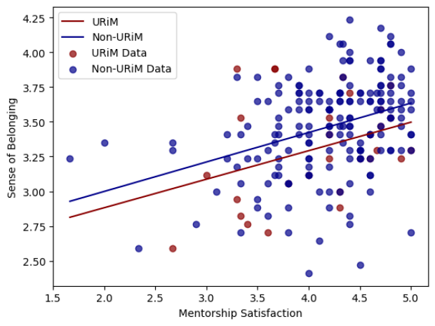
Figure 1. URiM students report a lower sense of belonging in medical school compared to non-URiM students.
Dr. Sophia Y. Wang
2023 - 2024 TMA Innovation Grant Awardee
PhacoTrainer: Artificial Intelligence Dashboard for Surgical Performance Feedback
Cataracts are a clouding of the natural lens of the eye which occurs with natural aging and causes vision loss. Cataract surgery is a highly delicate microsurgical operation wherein the natural lens of the eye is removed and replaced by a clear artificial lens implant, thereby curing cataract and restoring clear vision. Cataract surgery is the most commonly performed surgery in the United States. A primary goal of ophthalmology surgical training is to learn to perform cataract surgery skillfully.
Our PhacoTrainer project is focused on building an artificial intelligence (AI) powered dashboard for cataract surgery performance metrics, to enhance surgical training. We have developed AI models that can determine in each frame of routinely captured surgical video 1) what steps of cataract surgery are being performed, and 2) where the instruments and key anatomical landmarks are located. From this, we can also calculate metrics based on instrument and pupil motion, such as total path length, maximum velocity, and area covered of each instrument and anatomical landmark. We have shown that these performance metrics can distinguish between attending-performed vs trainee-performed surgeries, and that they correlate with expert human judges of performance using a well-established structured cataract performance rating scale. We are building a web application to which users can upload their routinely captured cataract surgical videos and receive AI-powered surgical metrics that allow them to track the time spent on each step of surgery, complication rates, and tool-motion based metrics.
The TMA innovation grant has enabled us to greatly expand the development of our application by implementing the newest AI models and motion-based performance metrics. In addition, we have recruited several ophthalmology residents to test our application to provide technical and holistic feedback. Studies are ongoing which will track and correlate the AI-performance metrics as residents progress in their training. Our goal is to improve cataract surgical training by providing trainees with granular and trackable performance metrics to guide their surgical development, ultimately enhancing patient outcomes.
Click here to read more about Dr. Wang's TMA Innovation Grant and about our other Grantees!
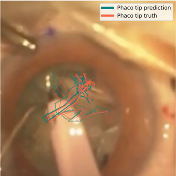
Figure 1. AI-identified instrument location compared to human-annotated instrument prediction. The blue line shows the location of the tip of the phacoemulsification probe over time during cataract surgery, as identified by the artificial intelligence model. The phacoemulsification probe is the key surgical instrument which breaks up and removes the cataract from the eye. The red line shows the same, but as annotated by a human. One can see that the identified location of the phacoemulsification probe tip is very close to the human-annotated location.
Dr. Amanda Rigas & Dr. Margaret Lin
2023 - 2024 TMA Innovation Grant Awardee
Reducing Interpretive Errors on Common Imaging Studies through Deliberate Practice and Mastery Learning
Our project involves integrating and validating principles of deliberate practice and “mastery” learning into Radiology image interpretation. We are utilizing STELLA (STanford Electronic Learning Library & Applications), a teaching file software platform created by Stanford radiologist Dr. Christopher Beaulieu, which can create searchable metadata and display radiological images from Stanford’s Picture Archive and Communication System (PACS). We will integrate quizzes and didactic information into STELLA to train first-year Radiology residents in critical imaging findings prior to taking independent call, including identification of pneumothorax, pneumoperitoneum, and misplaced support devices on adult xray. This open-source software has the potential to be used for other applications, including correlation with image rich information from other specialties such as Pathology, Radiation Oncology, Ophthalmology, Surgery, and Dermatology. Our ultimate goal is to deploy STELLA and mastery learning techniques to augment the training of any clinicians involved in image interpretation. More background on STELLA is available at: https://stella.stanford.edu.
Please reach out to Drs. Rigas, Lin, or Beaulieu if you would like to learn more or see a demo!
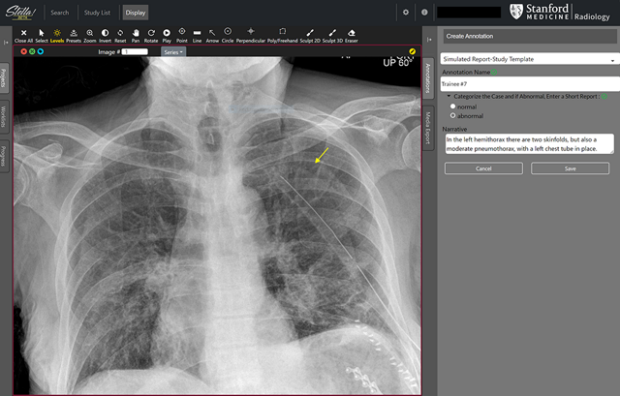
Figure 1. A screenshot of a teaching case in STELLA, with yellow annotation indicating the finding and narrative description in the side panel.