Skin
-
Lanugo
-
Normal Peeling
-
Icthyosis
-
Normal Skin Pigment
-
Albinism
-
Slate Grey Patches (Mongolian Spots)
-
Salmon Patch
-
"Stork Bite" Mark
-
Port Wine Stain
-
Hypopigmented Macule
-
Milia
-
Sebaceous Hyperplasia
-
Erythema Toxicum
-
Subcutaneous Fat Necrosis
-
Skin Irritation
-
Transient Neonatal Pustular Melanosis
-
Petechiae
-
Bruising
-
Cutis Marmorata Telangiectasia Congenita
-
Mottling (Cutis Marmorata)
-
Early Hemangioma
-
Congenital Hemangioma
-
Epidermal Nevus
-
Sebaceous Nevus
-
Junctional Melanocytic Nevus
-
Cafe Au Lait Spot
-
Cutis Aplasia
-
Sucking Blister
-
Skin Tags
-
Fingernail Scratches
-
Forceps Mark
-
Vacuum Mark
-
Scalp Electrode Site

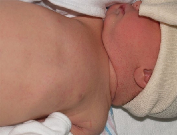
Lanugo
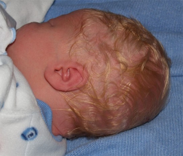
The downy hair seen over the shoulder is lanugo. Although this is present to a much greater degree in premature infants, term babies also have variable amounts of lanugo present at birth, as evidenced in this photo.
photo by Janelle Aby, MD
Normal Peeling
A dry, flaky, peeling appearance of the skin is very common in newborns. Although this can be distressing to parents, it does not need treatment and will spontaneously resolve.
photo by Janelle Aby, MD
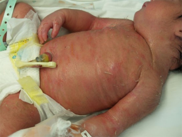
Normal Peeling
This is another example of a post-dates infant with dry, peeling skin. On the day of birth, the skin of this baby had a very leathery appearance. This photograph was taken on the second day, when the skin had dried out and the peeling was more visible. The red blotches visible on the chest, right forearm, and legs are erythema toxicum and are unassociated with the peeling.
photo by Janelle Aby, MD
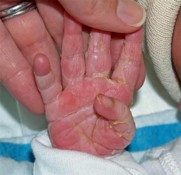
Normal Peeling
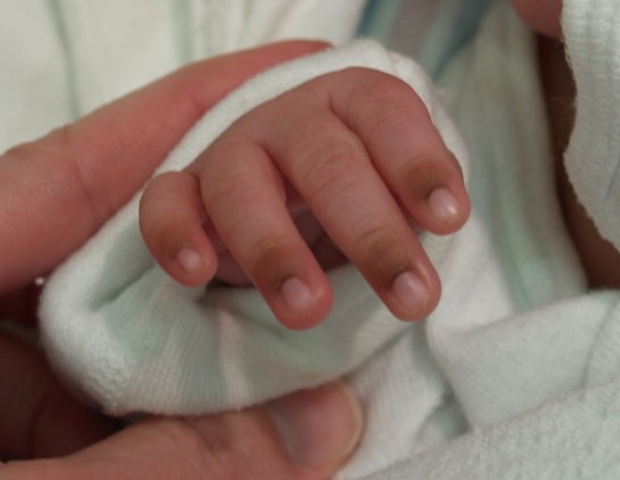
The hands and feet are especially common places to observe peeling. Often associated with infants who are post dates, many term babies exhibit this kind of peeling as well. In this patient, the skin and nails (look at the thumbnail) are stained yellow from the presence of meconium in utero. This photo was taken a few hours after birth, so the moist appearance of the skin is still visible. By the following day, the skin was more dry and the difference between the peeled and non-peeled areas of the hand was less visible.
photo by Janelle Aby, MD
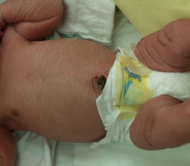
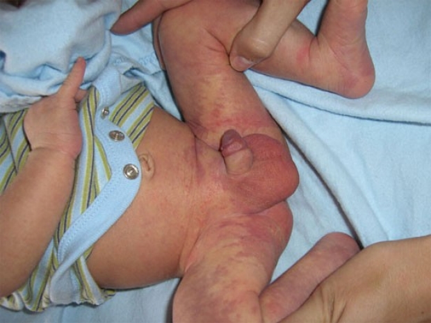
Icthyosis
In contrast, this type of peeling is not normal. Notice how the skin uder the peeled areas is red and fissured and how the fingers on the right hand appear edematous. This newborn had an uncomplicated pregnancy and delivery, but this skin appearance in the delivery room prompted transfer to the NICU for further evaluation. For this baby, Aquaphor was liberally applied to the skin, and the infant was kept in an isolette to minimize fluid losses.
photo by Janelle Aby, MD
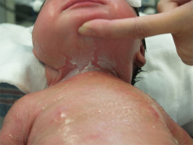
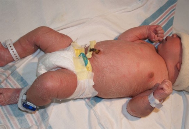
Icthyosis
This is the same infant as in the previous photo. Here, deeper fissuring can be seen in the area under the chin. Involvement of both pediatric dermatologists and geneticists is appropriate when dealing with congenital icthyosis.
photo by Janelle Aby, MD
Normal Skin Pigment
The increased pigment seen at the base of the nails is this African-American infant is entirely normal. When the parents' skin tone is dark, the overall skin tone of the baby will typically be much lighter than the parents at birth. In some areas, though, the increased melanin can be seen -- around the nails, over the helix of the external ear, around the umbilicus, and over the genitalia.
photo by Janelle Aby, MD
Albinism
This infant was born to African American parents. Although the pediatrician's physical examination was unremarkable except for the general hypopigmentation, eye findings consistent with oculocutaneous albinism were noted by the ophthalomologist. These included transillumination of the irises, blonde fundi and vessels running through the macula. Nystagmus was not present at birth, but may be present in this condition as visual acuity is often low.
photo by Janelle Aby, MD
Slate Grey Patches (Mongolian Spots)
These dark blue-grey lesions are most commonly seen in darker-skinned infants. The sacrum is the most commonly affected area. These lesions tend to fade over several years but may not completely disappear. No evaluation is needed. They can be easily differentiated from bruises by the absence of other colors associated with bruises -- red, purple, green, brown or yellow.
photo by Janelle Aby, MD
Slate Grey Patches (Mongolian Spots)
This baby has more intense and widespread lesions, but the diagnosis and management are the same.
photo by Janelle Aby, MD
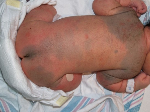
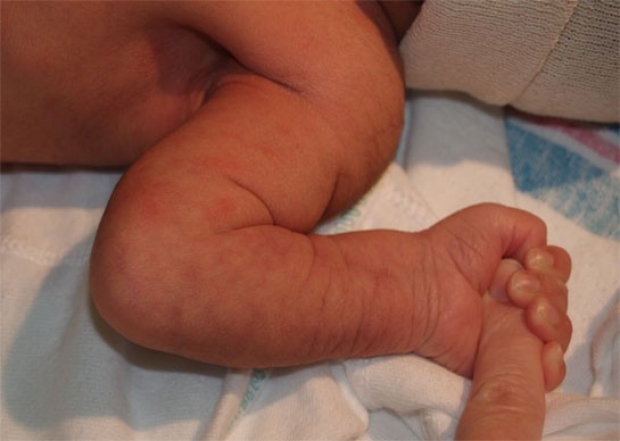
Slate Grey Patches (Mongolian Spots)
Slate grey patches can even involve an entire extremity. It is important to be able to distinguish the typical appearance of the patch even in an unusual location, so that trauma is not suspected. Patches involving a single leg or which are circumferential around both ankles have also been seen in our nursery. Documentation of these unusual birthmarks in the medical record is helpful.
photo by Janelle Aby, MD
Slate Grey Patches (Mongolian Spots)
This is another atypical example. These slate grey patches are typical in appearance, but unusual in location, as this finding is almost always observed on the back or extremities.
photo by Janelle Aby, MD

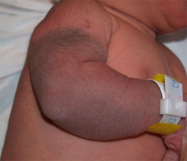
Slate Grey Patches (Mongolian Spots)
This photograph was taken approximately 6 hours after birth. In this case, the infant was initially thought to have sustained a bruise from delivery because this hand presented in a position on top of the head. The homogenous appearance and discrete borders, though, are inconsistent with a fresh bruise.
photo by Janelle Aby, MD
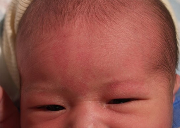
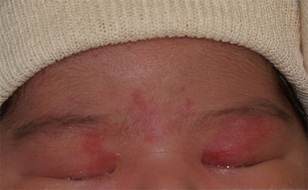
Salmon Patch
The pink patches in the middle of the forehead and over the left eye are salmon patches. Also known as nevus simplex or "angel kisses", these are a common capillary malformations that are present at birth. Eyelid spots generally fade over several months. Lesions on the glabella may take several years to resolve, and occasionally the outlines can be seen into adulthood, especially when the face is flushed.
photo by Janelle Aby, MD
Salmon Patch
This is another newborn with salmon patches. When lesions are present only on the eyelids, they are sometimes mistaken for bruising, but the examiner should realize that the eyelids are in a very protected position. Although the lids may be quite edematous, bruising in this location would be highly unusual, even in a baby with bruising elswhere on the face.
photo by Janelle Aby, MD
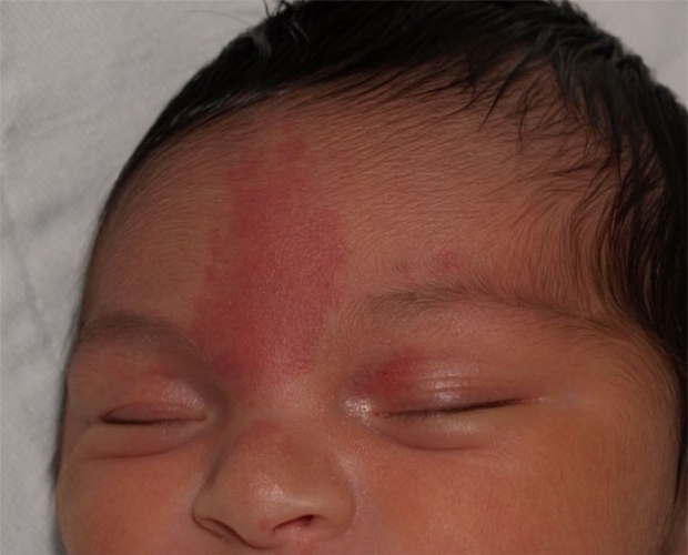
Salmon Patch
Here is a third example. In this patient, small spots can be seen over the eyelids and near the tip of the nose in addition to the large spot on the forehead.
photo by Janelle Aby, MD
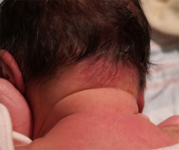
"Stork Bite" Mark
Salmon patches may also be found on the nape of the neck in newborns. Nicknamed "Stork Bite Marks", these lesions become less intense with time, but are frequently visible into adulthood.
photo by Janelle Aby, MD
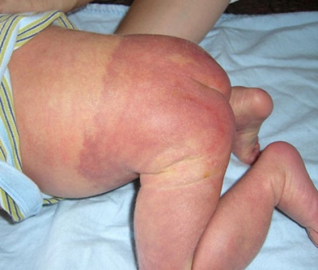
Port Wine Stain
In contrast, these congenital pink patches are port wine stains. They are typically more intense and purple-red in color than salmon patches. In some cases, as seen here, a port wine stain may affect a large surface area. The discoloration is not, of itself, a problem, but it may be a clue to an underlying condition. Port wine stains on the face may be associated with Sturge-Weber syndrome, and those on the extremities may be associated with Klippel-Trenaunay-Weber syndrome, in which overgrowth of an extremity may occur.
photo by Janelle Aby, MD
Port Wine Stain
Lesions that occur over the spine may also indicate an occult spinal dysraphism, so imaging should be considered. This is the same infant as in the previous photo. Although the lesion in this infant is not localized to the back, a spinal ultrasound was done; it was normal.
photo by provided by the parents
Hypopigmented Macule
Hypopigmented macules are typically very subtle, and benign, findings in the newborn. In this patient, the macule partially overlaps the slate-grey patch (9 o'clock position) on the upper thigh.
Many hypopigmented macules are transient, and are caused by abnormal local vasoconstriction, as in the patient above. In this case the lesion became more visible with gentle stroking of the skin, but otherwise was almost invisible.
If it is important to differentiate vasoconstriction from true hypopigmentation, a wood's lamp can be used. If an abnormality of pigment exists, the lesion will appear bright under a wood's lamp, while areas of abnormal vasoconstriction will appear the same as the surrounding skin.
Ash leaf spots, associated with tuberous sclerosis, are rare in newborns; usually they appear in children more than 5 years old.
photo by Janelle Aby, MD
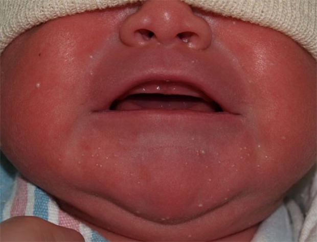
Milia
The white papules on this baby's chin and cheeks are milia. Milia are keratin filled epithethial cysts which occur in up to 40% of newborns. Spontaneous exfoliation and resolution is expected within a few weeks.
Parents will occasionally mistake these lesion for neonatal acne, but milia are present at birth and have no inflammatory component. Acne, even though caused by maternal hormones, does not generally appear until after 2 weeks of age.
photo by Janelle Aby, MD
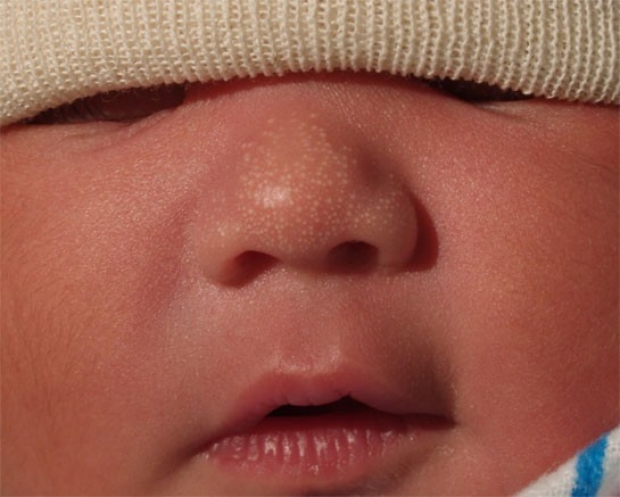
Sebaceous Hyperplasia
In contrast to milia, the raised lesions on the nose in this newborn are sebaceous hyperplasia. The lesions are more yellow than milia and are the result of maternal androgen exposure in utero.
Sometimes referred to as "the miniature puberty of the newborn," maternal hormone exposure may also cause vaginal withdrawl bleeding in infant girls and neonatal acne.
Sebaceous hyperplasia is a benign finding and spontaneously resolves with time. No evaluation is needed.
photo by Janelle Aby, MD
Erythema Toxicum
This is the "rash" most commonly observed in the nursery. Lesions generally start on day 1 or 2 and increase in number over the next several days, followed by spontaneous resolution in about a week. Even newborns who have hundreds of spots are not symptomatic and need no futher evaluation.
photo by Janelle Aby, MD
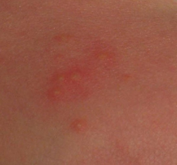
Erythema Toxicum
With a closer view, the typical lesion can be seen. A central, yellowish papule is surrounded by a halo of erythema.
photo by Janelle Aby, MD
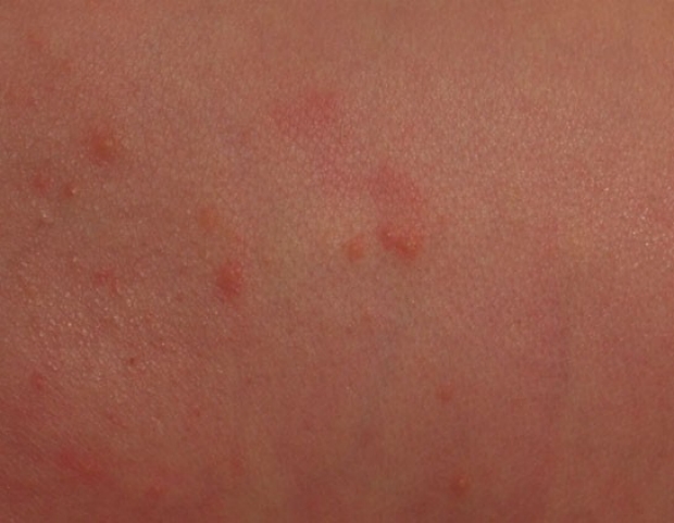
Erythema Toxicum
Another example of this ubiquitous rash. More than half of all newborns will have this to some degree.
photo by Janelle Aby, MD
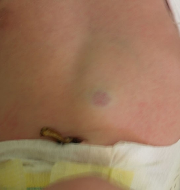
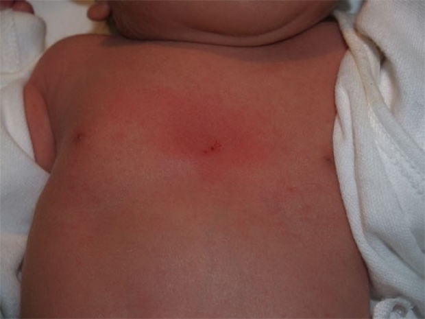
Subcutaneous Fat Necrosis
This red lesion is subcutaneous fat necrosis. On palpation, there is a firm nodule in the subcutaneous tissue under the area of redness that is freely mobile with respect to the bony structures underneath it.
Subcutaneous fat necrosis is more common in infants who have had difficult deliveries, cold stress, or perinatal asphyxia.
Lesions are typically asymptomatic and resolve spontaneously within several weeks, usually without scarring or atropy.
Infants with extensive lesions or with renal disease should have calcium levels followed once or twice weekly. Hypercalcemia associated with subcutaneous fat necrosis is rare, but is a potentially lethal complication.
photo by Janelle Aby, MD
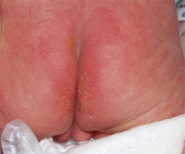
Skin Irritation
These raised yellow lesions are the result of irritated skin. Some newborns will have particularly sensitive skin, and may develop a "rash" of this type even in the absence of diarrhea or other known offending agents.
Symptomatic care and good diaper hygiene (with thorough drying before the diaper is replaced) is recommended.
photo by Janelle Aby, MD
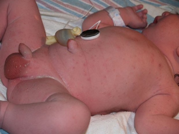
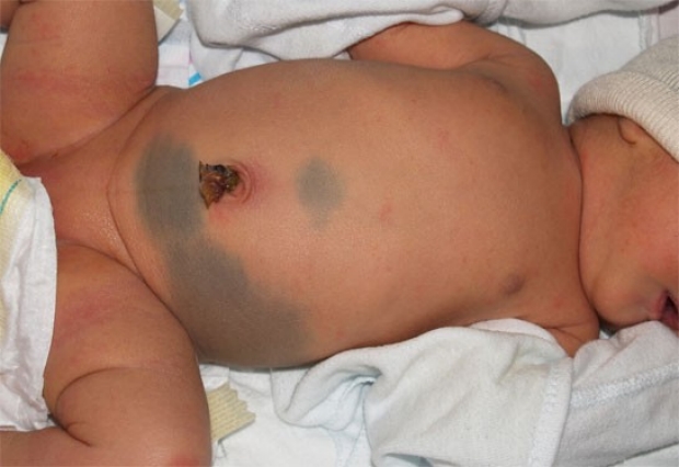
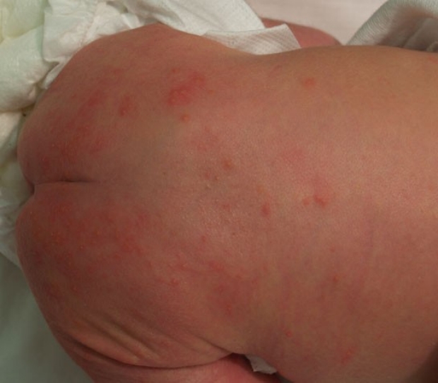
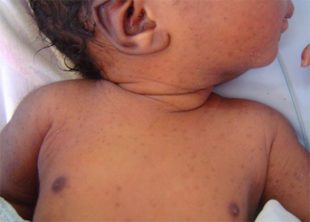
Transient Neonatal Pustular Melanosis
All the lesions seen here are consistent with this diagnosis. The hallmark of this rash is the hyperpigmented spots that remain (seen here on the chest) after the fragile pustules (seen on the scrotum and thigh) have resolved.
Because the rash starts in utero, lesions may be in any stage at birth.
Despite the anxiety-provoking appearance of a newborn covered in pustules in the delivery room, no evaluation is needed when non-inflammatory pustules occur in combination with hyperpigmented macules in an otherwise well infant.
photo by Janelle Aby, MD
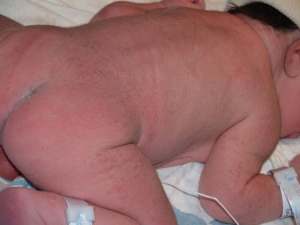
Transient Neonatal Pustular Melanosis
The back of the infant in the previous photo. Less pustules are noted here, but there are more hyperpigmented macules. These are expected to fade over several months. The pustules are fragile and last only a day or two.
photo by Janelle Aby, MD
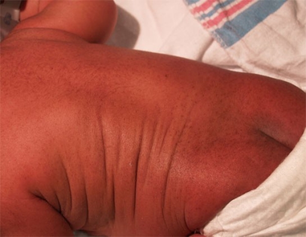
Transient Neonatal Pustular Melanosis
This infant was born with only the hyperpigmented macules present. Although the etiology is unknown, it has been observed that African Americans infants are more frequently affected with this condition, occurring in up to 4%. Spontaneous resolution is expected.
photo by Janelle Aby, MD

Transient Neonatal Pustular Melanosis
Here is a close-up of the lesions present in the previous photo.
photo by Janelle Aby, MD
Transient Neonatal Pustular Melanosis
This is another infant with the hyperpigmented macules typical of neonatal pustulare melanosis. In this photo, the "collarette of scale" that often surrounds the individual lesions can be appreciated (look carefully at the neck and the infant's left axilla/arm).
photo by Janelle Aby, MD
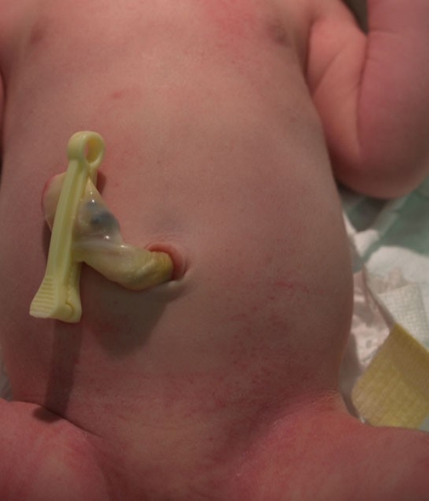
Petechiae
Petechiae were first noted in this infant in the groin, where they are most numerous. With a careful examination, spots can also be seen on the abdomen and left upper chest. While petechiae may be due to pressure during birth, widespread petechiae deserve some evaluation; a CBC and platlet count in this infant was normal.
photo by Janelle Aby, MD
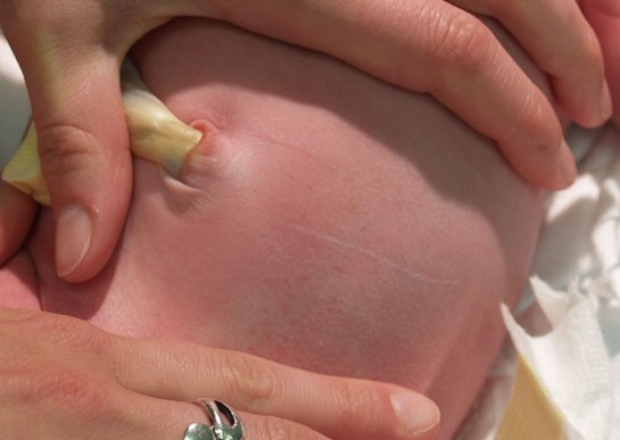
Petechiae
Blanching the abdomen reveals more petechiae than were appreciated on casual examination.
photo by Janelle Aby, MD
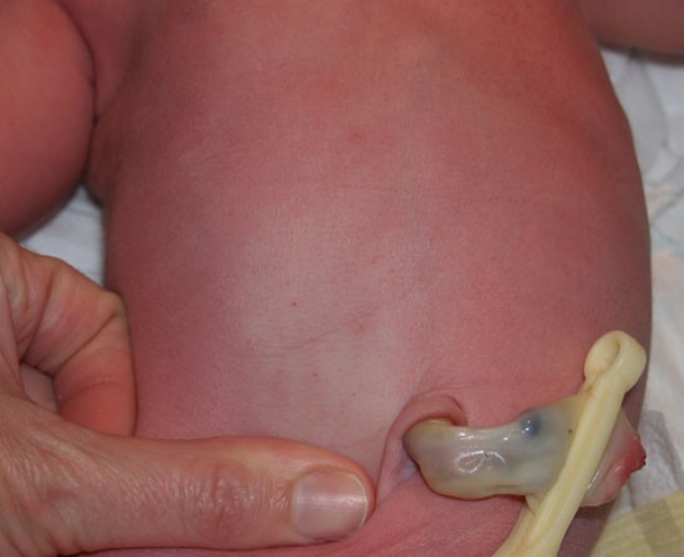
Petechiae
The same infant as in the previous photo, with more petechiae highlighted by blanching.
photo by Janelle Aby, MD
Petechiae
Another infant has petechiae primarily around the chin, neck, and upper chest. Given the relatively localized area affected in a newborn born vertex, no evaluation was done. These improved substantially over the next 2 days.
photo by Janelle Aby, MD
Bruising
This infant had a somewhat difficult delivery, and as a result, sustained bruises on the head and the left arm. This picture was taken on the third day of life. By this time, the bruises were significantly improved from the appearance at birth.
photo by Janelle Aby, MD
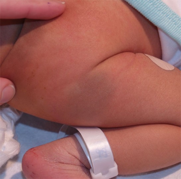
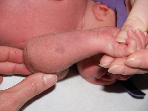
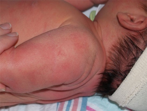
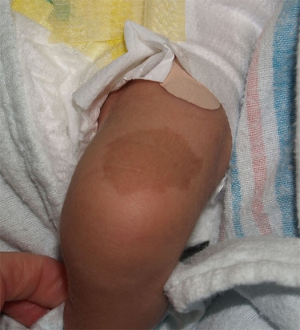
Cutis Marmorata Telangiectasia Congenita
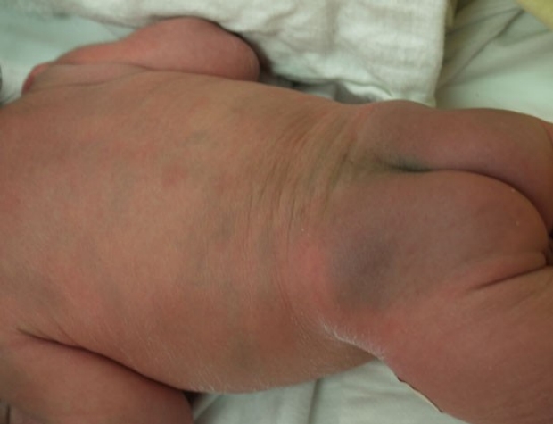
On the initial physical examination, this infant was thought to have sustained bruising of the right arm during delivery. On closer inspection, however, the discolored areas are also depressed, suggesting some underlying atrophy of the skin.
This is cutis marmorata tealngiectasia congenita, a rare congenital vascular malformation. The presentation seen here with one limb affected is most common. Associated defects may occur in up to 50% of affected patients, but the cutaneous lesions tend to improve with time.
photo by Janelle Aby, MD
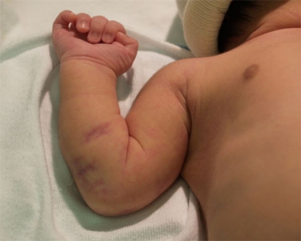
Cutis Marmorata Telangiectasia Congenita
In another patient, this dark purple lesion was noted on initial exam. Port wine stain and bruising were considered in the differential diagnosis, but the angulated shape made these possibilities less likely. In addition, there is a suggestion of dark erythema streaking from the lesion across the wrist.
See the following photo for more findings that helped lead to the diagnosis of cutis marmorata telangiectasia congenita.
photo by Janelle Aby, MD
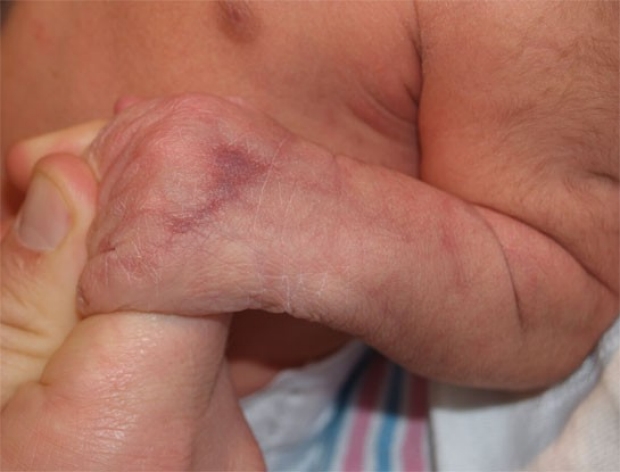
Cutis Marmorata Telangiectasia Congenita
When the underside of the forearm was examined, mottled streaks of erythema were noted.
Although mottling occurs commonly in newborns exposed to cool temperatures, it is transient (disappears with rewarming) and more generalized than this. Compare the mottled appearance of the forearm to the normal appearance of the upper arm to appreciate the difference.
This photo is a good example of how this condition got its name. Cutis marmorata is the term used for mottling of the skin; in this case, the mottling is of the congenital, telangiectatic variety.
Again, the skin lesions are expected to improve with time, but lesions should be followed for possible ulceration.
photo by Janelle Aby, MD

Mottling (Cutis Marmorata)
The lacy erythema present on the thigh of this newborn is mottling. Not to be confused with cutis marmorata telangiectasia congenita, cutis marmorata (mottling) is a transient and common finding in newborns. It is particular visible when the infant is cold and disappears with warming. The mottling in this photo is mild.
photo by Janelle Aby, MD
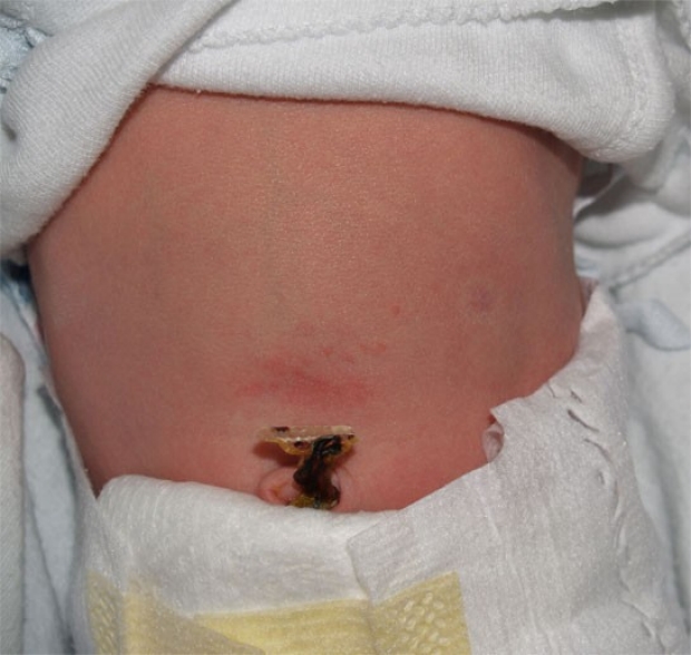
Early Hemangioma
In the newborn nursery, the raised, red appearance of a strawberry hemangioma is not usually seen. Instead, the precursors of these lesions are found. Although this photo is somewhat blurry, the lesion at this stage contains a central area of telangiectasia surrounded by a halo of pallor.
photo by Janelle Aby, MD
Early Hemangioma
This lesion is much more subtle and is completely flat, but the circular area of pallor with central vascular markings can still be seen on the upper arm of this newborn.
photo by Janelle Aby, MD
Early Hemangioma
This example is a little more noticeable than the previous one, but is still a subtle finding. The small, circular, pale area on the left side of the baby's abdomen (right side of the photograph) is the hemangioma. In this position, no problems would be expected. Lesions located in an area of chronic irritation (diaper area) or near vital structures (eye) can be problematic and may require treatment when they grow and become raised, but typically the best cosmetic result is obtained if the lesion is left to grow and involute spontaneously.
photo by Janelle Aby, MD
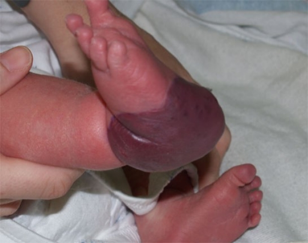
Congenital Hemangioma
A much more unusual presentation of hemangioma is shown here. This vascular lesion on the heel was present at birth. Initially, this was thought to possibly be a hemangioendothelioma, a lesion with malignant potential. Eventually however, that diagnosis was excluded. This particular infant did not develop Kasabach-Merritt syndrome, though that was a concern, and the lesion began spontaneously involuting after about 6 months of age.
photo by Janelle Aby, MD
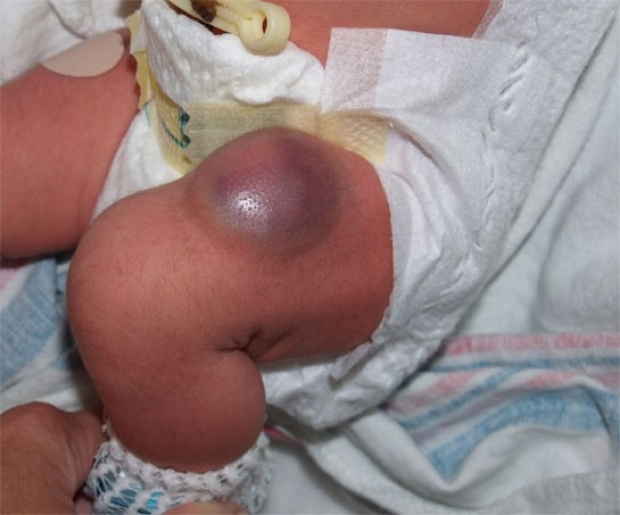
Congenital Hemangioma
This is another fairly large congenital hemangioma. In this case, the hemangioma appears to have more deep, rather than superficial, involvement. Again, unless complications arise, the best course of action seems to be watchful waiting.
photo by Janelle Aby, MD
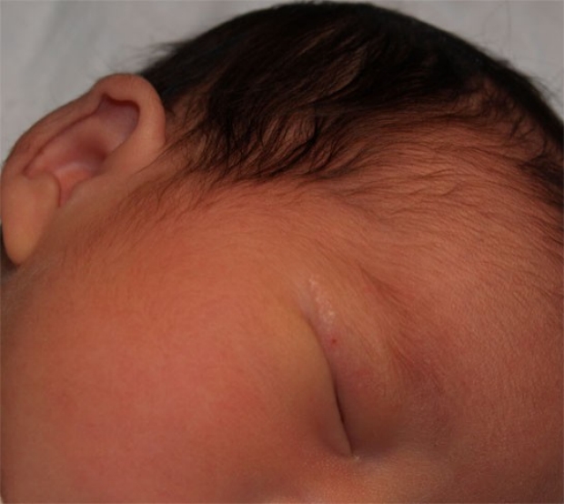
Epidermal Nevus
This lesion was present at birth. Initially, there was concern that this was a group of vesicles, but on careful examination, the lesion is firm and papular without any evidence of inflammation. This is consistent with an epidermal nevus. With time, epidermal nevi usually become more wart-like and scaly. Patients with epidermal nevi may have associated CNS, bone, and eye abnormalities, but this is more likely in those with extensive lesions. This baby was otherwise well.
photo by Janelle Aby, MD
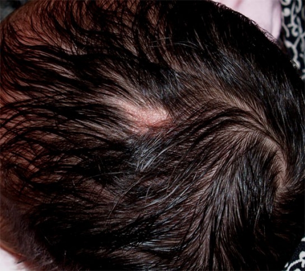
Sebaceous Nevus
A sebaceous nevus (also known as sebaceous nevus of Jadassohn) is a yellow-orange, waxy, pebbly lesion present on the face or scalp of some newborns. Hair follicles are not present within the lesion itself, but lesions on the scalp may be covered over by surrounding hair, so careful examination is important.
photo by Janelle Aby, MD
Sebaceous Nevus
Here is another example of sebaceous nevus on the lateral margin of the eyelid. The significance of this finding is primarily related to the fact that it is sensitive to the androgens produced during puberty, causing the lesion to become larger and more wart-like, so elective removal may be considered at that time. Almost all of the changes that occur within these lesions are benign, but there are a few reports of basal cell carcinomas in older adults, so observation throughout life is important.
photo by Janelle Aby, MD
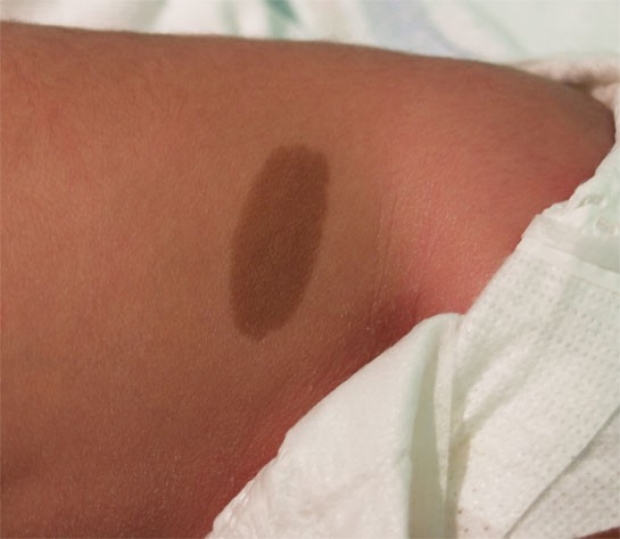
Junctional Melanocytic Nevus
Here is a typical appearance of a junctional melanocytic nevus. The lesion is completely flat and is medium to dark brown in color. It may be present at birth, as it was in this infant. It may become slightly raised as the infant grows and may become a compound nevus if intradermal melanocytes develop. It is considered a benign lesion.
photo by Janelle Aby, MD
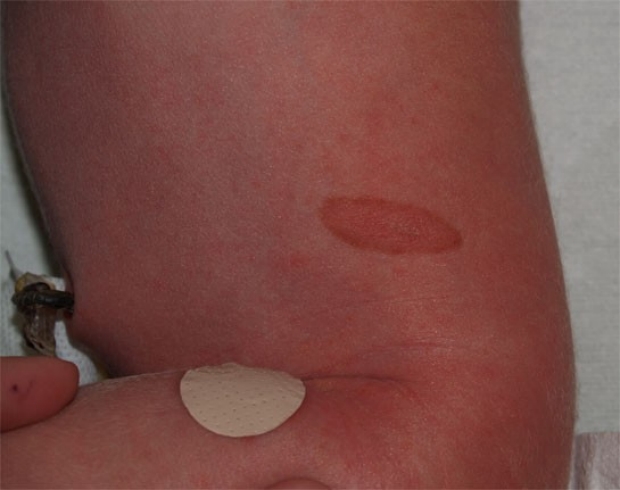
Junctional Melanocytic Nevus
This is a less typical appearance for a congenital melanocytic nevus. On initial examination, the central portion of this lesion appeared to be peeling (minimal peeling can be seen in this photograph). It is also more common to have a dark center with a light rim than it is to have this "halo" appearance.
photo by Janelle Aby, MD
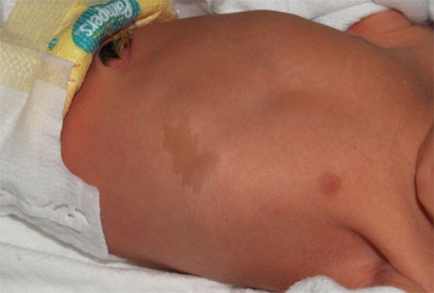
Cafe Au Lait Spot
Cafe au lait spots are lighter in color than melanocytic nevi, but they also may be congenital macules. They are caused by an increased amount of melanin in both melanocytes and epidermal cells, and may increase in number with age. Most children with cafe au lait spots do not have neurofibromatosis, but the presence of six or more of these spots that are larger than 0.5cm diameter is considered to be a major clue to the diagnosis.
photo by Janelle Aby, MD
Cafe Au Lait Spot
Here is another example of cafe au lait spot. Again in this patient, the spot was an isolated physical finding, so no further evaluation was necessary.
photo by Janelle Aby, MD
Cutis Aplasia
On examination of this infant, there were 3 areas of abnormality -- the nodule under the left eye, the red "excoriated" area lateral to the nodule, and the hairless, circular lesion anterior to the ear. The etiology of the nodule is unknown at this time, but the other two areas were felt to be consistent with cutis aplasia. Cutis aplasia is often thought of in association with trisomy 13, but it can also be an isolated finding in an otherwise well newborn, as in this case.
photo by Janelle Aby, MD
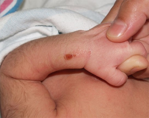
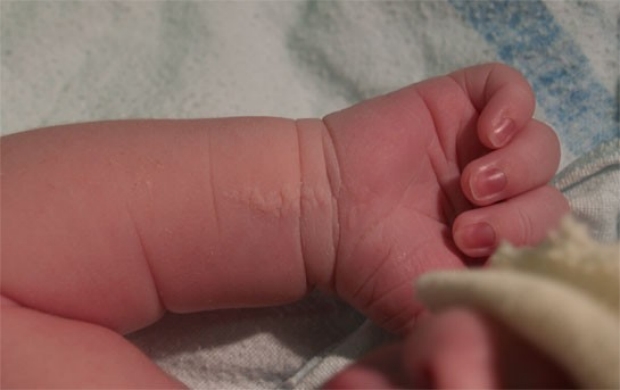
Sucking Blister
This lesion is fairly typical of the appearance of a sucking blister at birth. While the blister created by the infant sucking on his extremity in the womb may still be intact at the time of delivery, often it appears as a flat, scabbed, healing area (as shown here). Sucking blisters are solitary lesions that occur only in areas accessible to the infant's mouth. They are benign and resolve spontaneously. The appearance and location of the lesion is usually sufficient for diagnosis, but if the infant is observed sucking on the affected area, the diagnosis is certain.
photo by Janelle Aby, MD
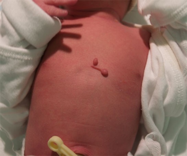
Skin Tags
These tags are rather typical in appearance, but in a highly unusual location. Both were pedunculated tags with very thin stalks, although the one on the baby's right is much longer. Because of the narrow diameter of the stalk, the decision was made to clip these with scissors instead of tying them off.
photo by Janelle Aby, MD
Skin Tags
A sharp, sterile scissors was used to remove the two tags. This photo, taken immediately after the procedure, shows an excellent result. There was no bleeding from the longer tag on the baby's right side; there was very slight bleeding from the spot on the baby's left, but after pressure was briefly applied, it quickly stopped.
photo by Janelle Aby, MD
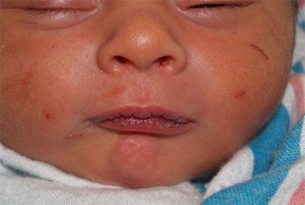
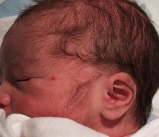
Fingernail Scratches
When babies are born, the nails are frequently rather jagged and sharp. Because normal behavior for a newborn includes bringing fists and hands up to the face, self-inflicted fingernail scratches are not uncommon.
In this infant, two linear scratches can be clearly seen on the patient's left cheek, but there are also several other subtle scratches visible (one under the right eye, one on the right cheek, one under the right nares, and one inside the left nares.
In an effort to prevent scratches, the hands of a newborn are frequently covered with long sleeves, mittens or socks. Fingernails can also be carefully trimmed with a nail file or clippers (a feat most easily accomplished when the baby is asleep).
photo by Janelle Aby, MD
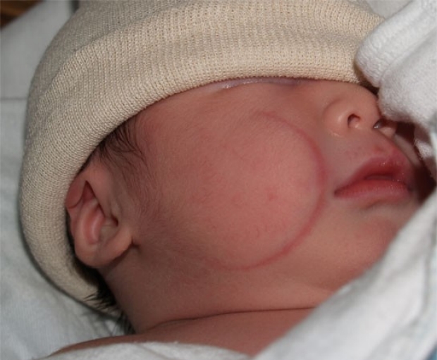
Forceps Mark
The semi-circular red mark on this infant's right cheek is a forceps mark. When forceps are needed to assist with a delivery, this type of superficial red mark can occasionally been seen on the sides of the infant's face. In most cases, the marks are small (<2cm) erythematous streaks. These marks have no consequence and will spontaneously resolve. In rare cases when the skin is abraded, antibiotic ointment may facilitate healing.
photo by Janelle Aby, MD
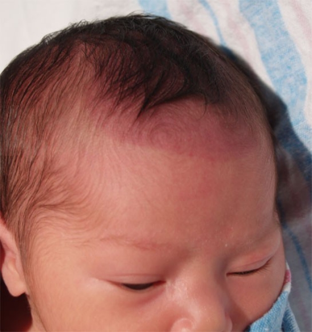
Vacuum Mark
Vacuum extraction can also leave marks on the infant's head. Typically, the bruising is similar to that which occurs normally during the process of delivery except for the fact that it is well circumscribed. In some cases, however, the bruising can be more severe with associated blisters or sloughing of the skin, or with underlying cephalohematoma or subglaeal hemorrhage. In this infant, the bruising is fairly mild, but can be easily seen because it extends beyond the hairline in the front. This finding spontaneously resolved.
photo by Janelle Aby, MD
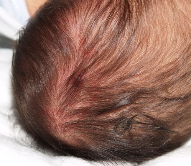
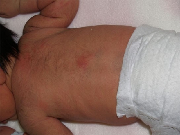
Scalp Electrode Site
When a scalp electrode is used for internal monitoring prior to delivery, a small circular scab can often be seen at the site where the monitor was. In this child, the scalp electrode site is the slightly red area with central darkness in the middle of the photograph, just to the right of the part in the hair. This amount of erythema is consistent with normal healing, but as this site is a break in the skin, infection in this area is possible (The lighter red spots on the scalp are consistent with bruising).
photo by Janelle Aby, MD