Eyes
-
Eyelid Edema
-
Dysconjugate Eye Movements
-
Dacrocystoceles
-
Dacrocystocele
-
Dacrostenosis
-
Subconjunctival Hemorrhage
-
Iris Cyst
-
Normal Eye
-
Injected Eye
-
Peripupillary Vascularity
-
Gonococcal Conjunctivitis
-
Congenital Glaucoma
-
Peter's Anomaly
-
Congenital Cataract
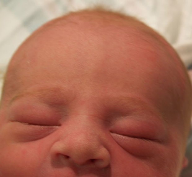
Eyelid Edema
Most infants exhibit some degree of eyelid edema after birth. The puffiness may make it seem that the infant has difficulty opening one or both eyes, but with a gentle examination, the eye can be easily evaluated. Edema resolves over the first few days of life.
photo by Janelle Aby, MD
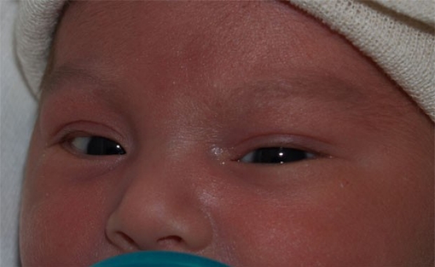
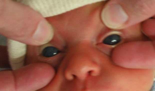
Dysconjugate Eye Movements
During the first few months of life, newborns will frequently have dysconjugate eye movements, where the eyes appear to move independently. Eyes may transiently appear crossed or divergent. This phenomena is particularly noticeable when the infant is falling asleep or being woken from sleep. If the dysconjugate movement is fixed (one eye is always out, or always in, relative to the other), a pediatric ophthalmologist should be consulted. But if the movements are transient, it is a normal finding and will spontaneously resolve.
photo by Janelle Aby, MD
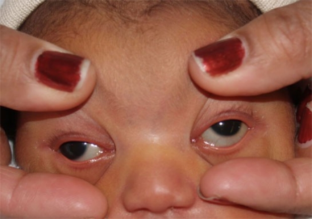
Dysconjugate Eye Movements
Here is another example of dysconjugate eye movements. This infant was sound asleep when the lids were gently opened for the photo. When awake, the infant could easily fix and focus on the mother's face.
photo by Janelle Aby, MD
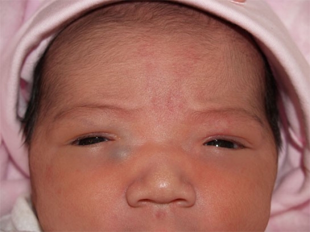
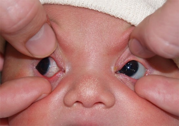
Dacrocystoceles
This infant was noted at birth to have some bluish nodules inferior to the medial canthi of both eyes, widely spaced eyes (hypertelorism), and a flat nasal bridge. The nodules are dacrocystoceles. They are caused by obstructions at both superior and inferior ends of the nasolacrimal duct. Though relatively small externally, these lesions almost completely obstructed the nasal passages. For this reason, internal nasal evaluation is recommended when the dacrocystoceles are bilateral, even when the patient is otherwise asymptomatic (as this child was).
photo by Janelle Aby, MD
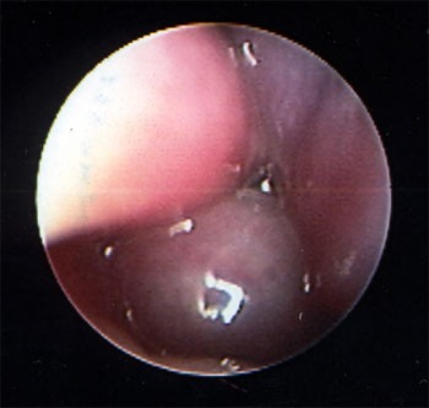
Dacrocystocele
An intra-operative photo shows the degree of obstruction of the nasal passage on the right side. The pink structure on the upper left is the turbinate, and the bluish mass at the bottom of the photo is the dacrocystocele. The small dark spot in the center was the area available for breathing.
photo by Anna Messner, MD
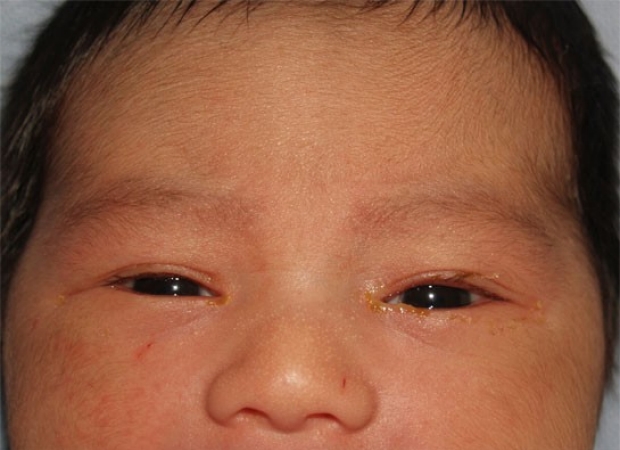
Dacrostenosis
Here is an example of dacrostenosis. In this case, both eyes are affected, although there is slightly more discharge around the left eye than the right. The sclera of both eyes are clear, and there are no other signs of infection. For about half of affected patients, this will resolve within the first week of life. For the other half, spontaneous resolution is expected in several weeks to a few months.
photo by Janelle Aby, MD
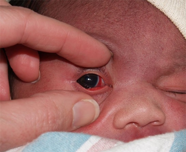
Subconjunctival Hemorrhage
Subconjunctival hemorrhage is a frequent finding in normal newborns. It results from the breakage of small vessels during the pressure of delivery. The red area may be large or small but is always confined to the limits of the sclera. It is asymptomatic, does not affect vision, and spontaneously resolves in several days. Note also the numerous petechiae on the forehead. This infant had significant facial bruising as well as the eye finding.
photo by Janelle Aby, MD
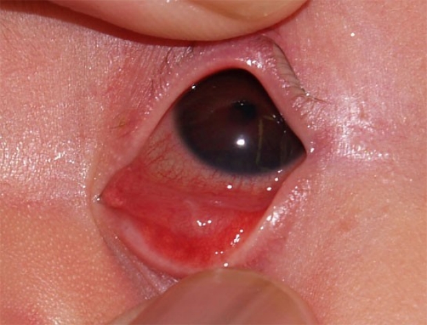
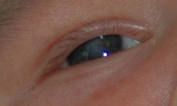
Iris Cyst
This infant had a normal physical exam at birth. By about 20 hours of life, there was a reddish/blue discoloration of the eyelid and redness in the left eye, the sclera was markedly injected, there was a small amount of discharge in the upper lashes, and the retinal light reflex was not visible. With careful inspection, a reddish discoloration over the iris (which gives the normally bluish iris a brown tint), and an opacity within the pupil can be seen. These findings were believed to be due to an iris cyst, an extremely rare finding. Interestingly, the cyst spontaneously resoved over a few months.
photo by Janelle Aby, MD
Normal Eye
For comparison, this is the completely normal right eye of the same infant. Note the lack of injection in the sclera, the normal greyish blue iris and the clear pupil. A retinal reflex was easily obtained in this eye.
photo by Janelle Aby, MD
Injected Eye
This is another newborn with unilateral conjunctival injection. On initial examination, the right pupil was about 2mm in diameter and the normal left pupil was 5-6mm. A retinal reflex was not present on the right side. This photo was taken after dilating drops were placed. Some dilation of the right pupil is noted, but it was not equal with the left, and still the retina could not be well seen. The differential diagnosis includes tumor and vascular abnormalities. An ultrasound of the eye was done to evaluate this finding further.
photo by Janelle Aby, MD
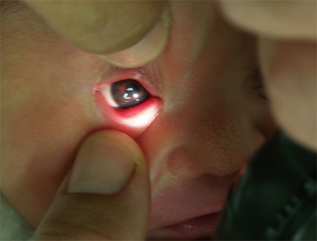
Peripupillary Vascularity
This is the same infant shown in the previous photo. Here the light from the ophthalmoscope is shining obliquely across the front of the eye. Without any special equipment, a red tinge (described by the ophthalmologist as a lacy network of peripupillary vessels) can be appreciated at the rim of the pupil. When the other eye was evaluated in this way, no red color was seen.
photo by Janelle Aby, MD

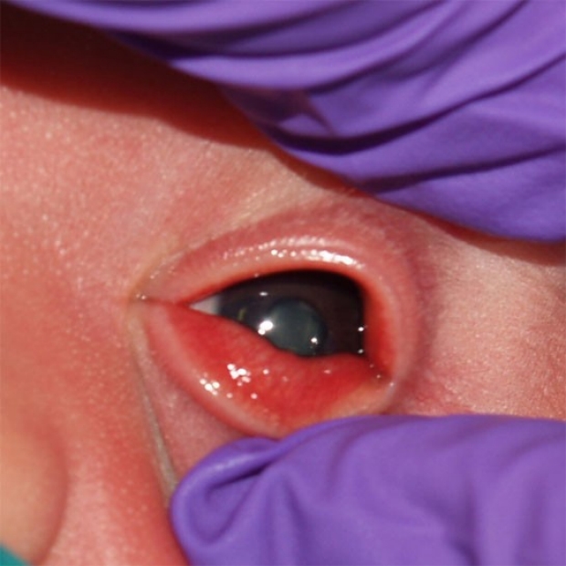
Gonococcal Conjunctivitis
When gonococcal conjunctivitis occurs, it typically presents in the first few days of life with copius, purulent discharge in the eyes. In this infant, the marked edema of the eyelids was the first symptom noted, but with just slight pressure on the lids, purulent material oozed out (seen here). Unlike the typical "pink eye" conjuncitivitis that occurs in older children, gonococcal conjunctivitis is an ophthalmologic emergency. Because the bacteria can erode through an intact cornea, treatment is very aggressive and includes systemic intravenous antibiotics, frequent eye washes, and monitoring in a neonatal intensive care unit. Fortunately, this diagnosis is very uncommon in places where prophylactic antibiotic eye ointment is used at birth.
photo by Janelle Aby, MD
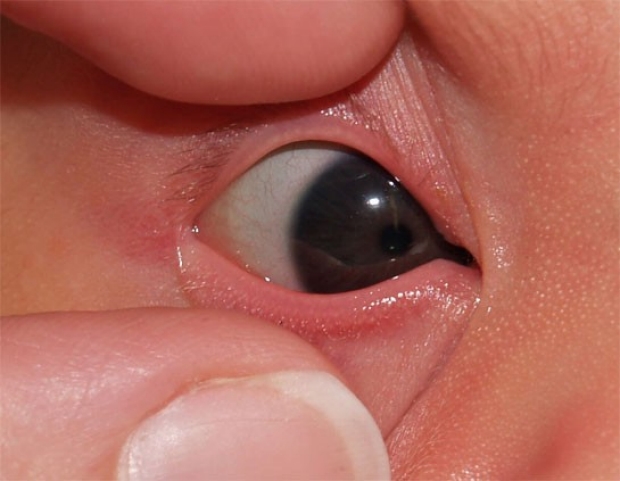
Congenital Glaucoma
This infant presented with hazy bilateral corneal opacities on the initial newborn exam. Congenital glaucoma was the underlying cause.
photo by Janelle Aby, MD
Peter's Anomaly
This infant has haziness of the central cornea visible without special equipment. On evaluation with an ophthalmoscope, retinal reflexes could not be seen due to this opacity. Although the opacity is central and could be mistaken for opacity of the lens, a view from a more oblique angle showed that the cloudiness extended over parts of the iris as well.
photo by Randall Young, MD
Congenital Cataract
In contrast to the previous photo, the opacities here occur behind the pupil, as the pupil is easily and clearly seen along its entire circumference. Again, the opacity can be appreciated without any special equipment (a good reminder to assess the eyes visually even if an ophthalmoscope is not readily available). A retinal light reflex could not be obtained. Congenital cataracts require early intervention to preserve sight, so immediate referral to a pediatric ophthalmologist is indicated. It is important to remember that cataracts that are less dense may not be visibly cloudy, so any time retinal reflexes cannot be obtained on exam, even if the eye appears grossly normal, referral is required.
photo by Janelle Aby, MD