Extremities
-
Normal Hand Crease
-
Transverse Palmar Crease
-
Polydactyly
-
Postaxial Polydactyly
-
Preaxial Polydactyly
-
Syndactyly of the Toes
-
Syndactyly
-
Polysyndactyly
-
Clinodactyly
-
Finger Hypoplasia
-
Toe Hypoplasia
-
Thumb Aplasia
-
Overlapping Toes
-
Positional Deformity of the Foot
-
Club Feet
-
Edema of the Feet
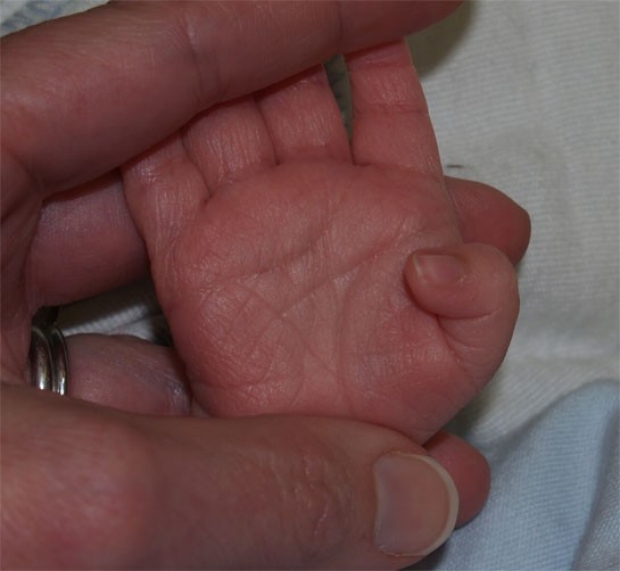
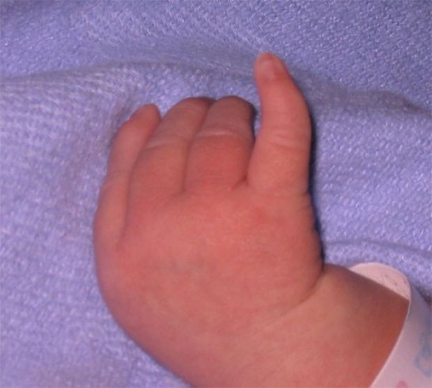
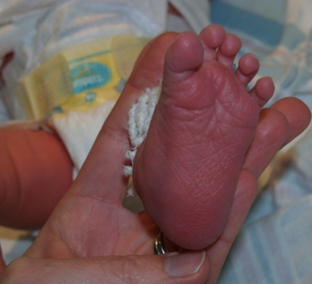
Normal Hand Crease
Most newborns have two major creases on the palm, neither of which completely extend from one side of the palm to the other.
photo by Janelle Aby, MD
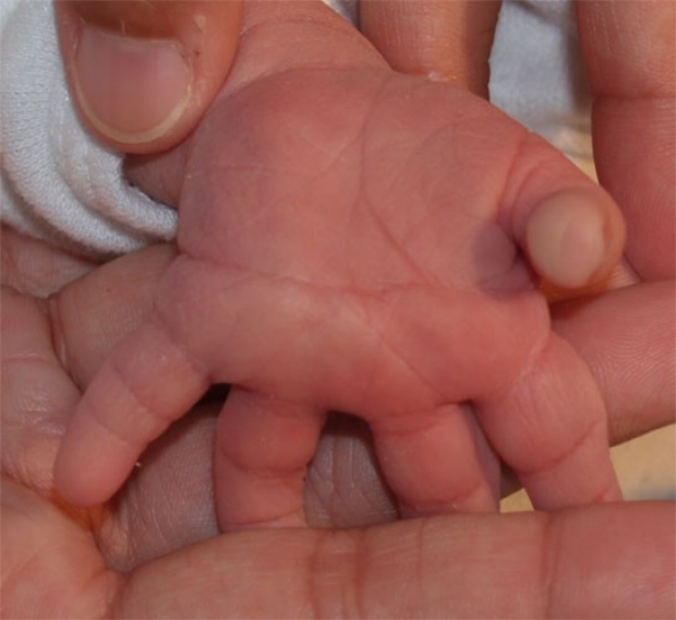
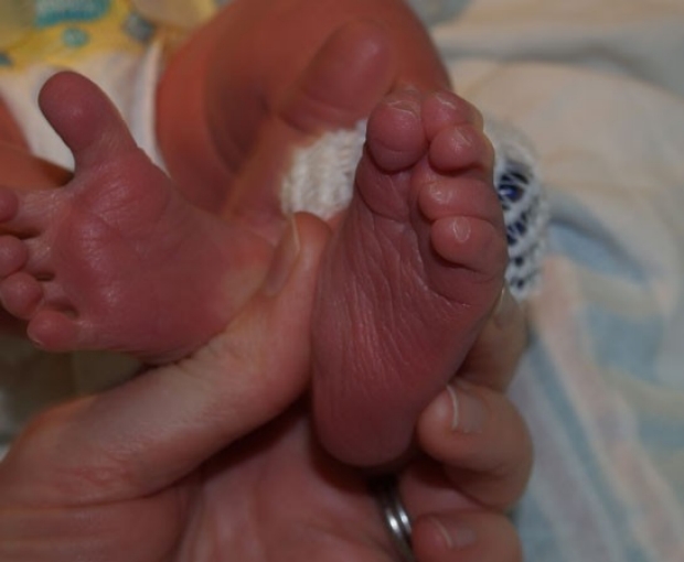
Transverse Palmar Crease
A common variant, found in approximately 5% of newborns, a transverse palmar crease is frequently inherited as a familial trait. Although single palmar creases are also associated with Down's syndrome and other genetic disorders, the absence of other abnormalities on physical exam should reassure the examiner that no further evaluation is necessary.
photo by Janelle Aby, MD
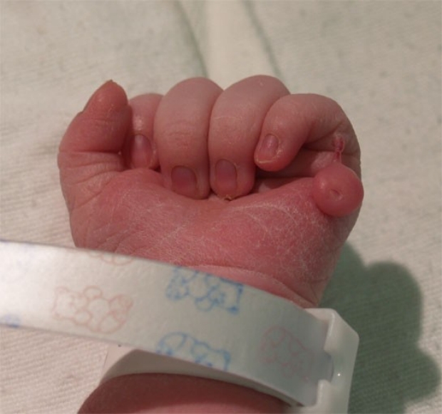
Polydactyly
Post-axial polydactyly is the most common variety. The extra digit may seem almost fully formed or may be attached only by a thin fleshy stalk. This is typically an isolated finding. A positive family history is often obtained.
photo by Janelle Aby, MD
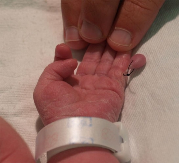
Polydactyly
In cases where the attachment of the extra digit is very thin, the appendage can be successfully tied off with a silk suture. After allowing some time for hemostasis to occur under the ligature, we usually snip off the appendage with a sterile scissors. The suture will either fall off spontaneously, or it may be removed in a few days.
photo by Janelle Aby, MD
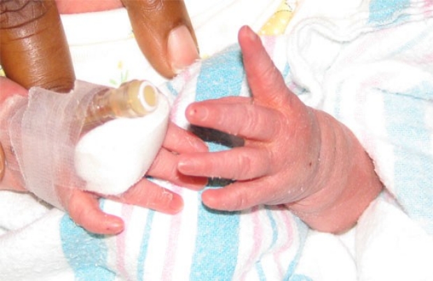
Postaxial Polydactyly
Another example of postaxial polydactyly. In this case the polydactyly is bilateral and the extra digits are fully formed.
photo by Janelle Aby, MD

Preaxial Polydactyly
This infant has polydactyly of the thumb as an isolated physical finding. There was no family history and the finding was unilateral. Preaxial polydactyly is much less common (and more likely to be related to an underlying medical condition) than the postaxial variety. Surgical correction is generally delayed for several months to lessen the risk of general anesthesia.
photo by Janelle Aby, MD
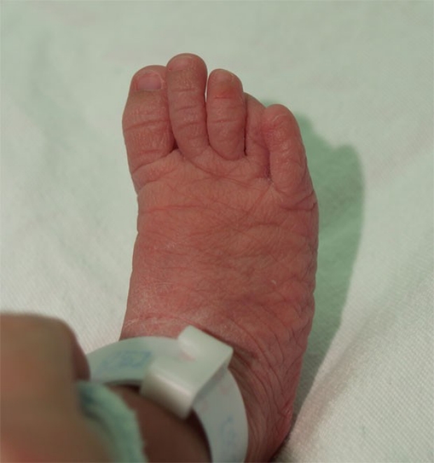
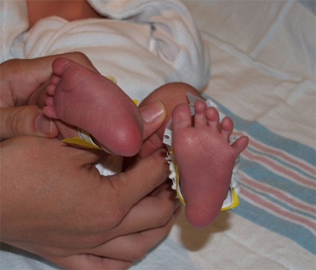
Syndactyly of the Toes
This type of mild syndactyly is found occasionally on the physical examination. Again, this is a subtle finding. If the exam is not a careful one, this can easily be missed since both digits are otherwise normally formed. This is usually an isolated finding, and in this location, has no impact on function.
photo by David A Clark, MD

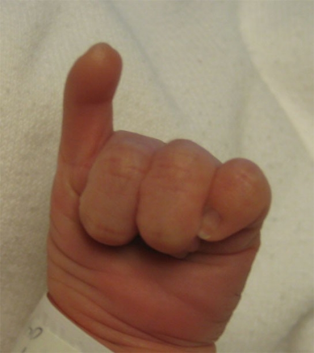
Syndactyly
In this case, the syndactyly is severe. All four of the digits on the right were completely fused together (notice the four fused nail beds). The thumb was normally formed and can be seen to the right of the abnormal area. This was a unilateral, isolated finding in an otherwise well newborn; amniotic bands were thought to have caused this problem.
photo by Janelle Aby, MD
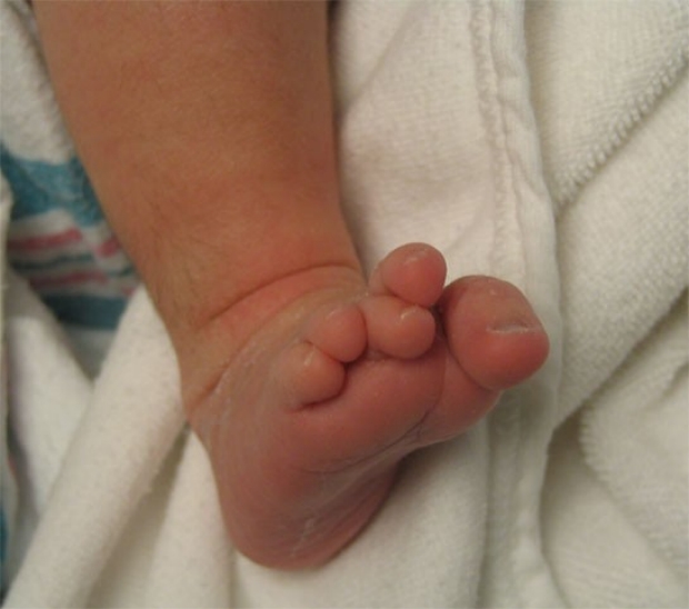
Polysyndactyly
When the digits are fused in addition to being too numerous, the term polysyndactyly is used. Careful examination is important to identify conditions such as this. If the socks (or mittens) are not removed, this finding can be easily missed. While this is not expected to cause a significant functional problem, cosmesis can be of concern as the child gets older.
photo by Janelle Aby, MD
Polysyndactyly
Here again polysyndactyly is present. In this case, the fifth and sixth digits are completely fused to each other. With careful inspection, a partial syndactyly of the second and third toes is also seen.
photo by Henry Lee, MD

Clinodactyly
A slight medial incurvation of the fifth finger is a frequent finding on the newborn exam. It is often inherited as a familial trait. Although it can be associated with several genetic disorders, including Down's syndrome, the absence of other physical findings will confirm that it is a benign, isolated finding. In this case, no further evaluation is necessary.
photo by Janelle Aby, MD
Clinodactyly
This infant has a more pronounced curvature. Again, this was an isolated bilateral physical finding. As seen in the next photo, this appearance was not a surprise for the mother, who had this trait as well.
photo by Janelle Aby, MD
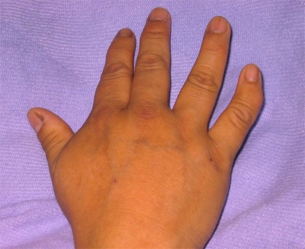
Clinodactyly
Here is the right hand of the mother of the infant in the previous photo. Both mother and baby were healthy -- this is a benign finding when isolated.
photo by Janelle Aby, MD
On first inspection, this infant's hands may appear normal, but on closer inspection, the fifth fingers bilaterally appear hypoplastic, and there is a complete absence of the nail on the infant's left side.
photo by Pete Pellegrino, MD
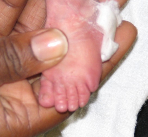
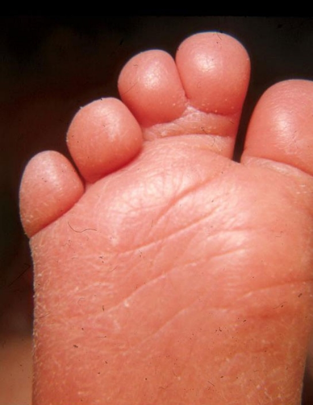
This is the same patient as the previous photo. Here, hypoplasia of the fifth toe (and nail bed) is apparent. This will not create a functional problem, but these findings did lead to an underlying diagnosis of Coffin-Siris syndrome.
photo by Pete Pellegrino, MD
Whenever digits are missing, underlying genetic or chromosomal diagnoses should be considered. Amniotic bands could also be responsible for absence of a digit, but in those children, the physical examination should be otherwise normal.
photo by Pete Pellegrino, MD
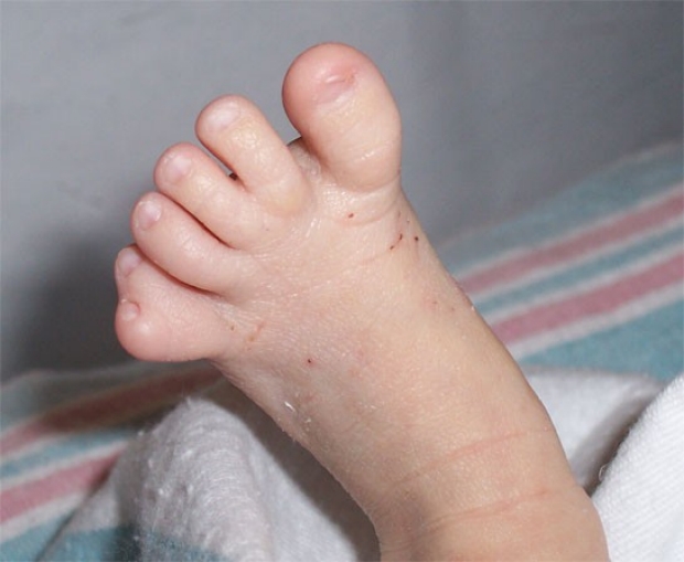
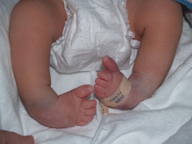
Some newborns will have toes that seem to overlap one another. At times, this is related to clinodactyly of one of the digits; at times, it is a positional deformity. Either way, it is considered a minor deformity. As an isolated finding, this is of no significance.
photo by Janelle Aby, MD
This infant has more marked overlapping of the toes. This child did have an underlying genetic diagnosis, but there were other abnormal physical findings present.
photo by Pete Pellegrino, MD
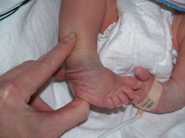
This infant appears to have a deformity of the left foot. Although bony deformities can occur, positional deformities are much more common. The following photos will show that this position is flexible and is therefore due to in utero position. Spontaneous resolution is expected.
photo by Janelle Aby, MD
A closer view shows how the sole appears to be everted.
photo by Janelle Aby, MD
With gentle pressure on the medial malleolus and the lateral aspect of the sole, the foot can be easily brought into a neutral position.
photo by Janelle Aby, MD
Another example of a positional deformity. In this case, the foot is dorsiflexed to such a degree that it is almost flat against the lower leg.
photo by Janelle Aby, MD

Here again, the foot can easily be brought back to a neutral position. A concavity due to the unusual position can be seen near the ankle, but this will resolve as the infant has full range of motion of the foot. in some cases, physical therapy that involves stretching and range of motion exercises can help speed progress.
photo by Janelle Aby, MD
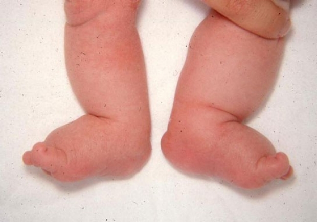
Bilateral club foot is noted in this infant. The feet are plantar-flexed and inverted. Because this is a bony deformity, this position is rigid.
photo by Janelle Aby, MD
With an attempt to bring the foot to neutral position, the rigidity of the ankle is apparent. Although the skin is sliding and wrinkling, the actual position of the foot is not changed. The dark discoloration around the ankles are slate grey patches (mongolian spots), not bruises.
photo by Janelle Aby, MD
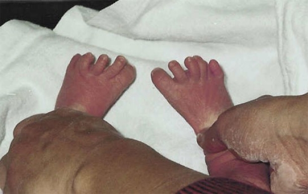
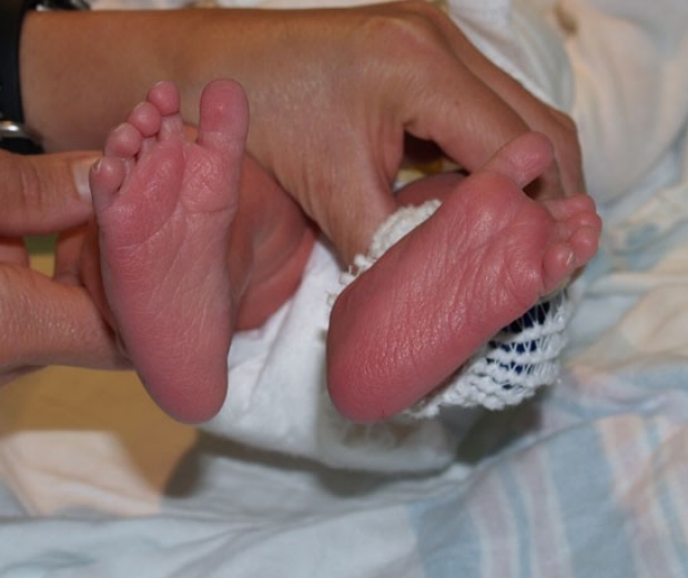
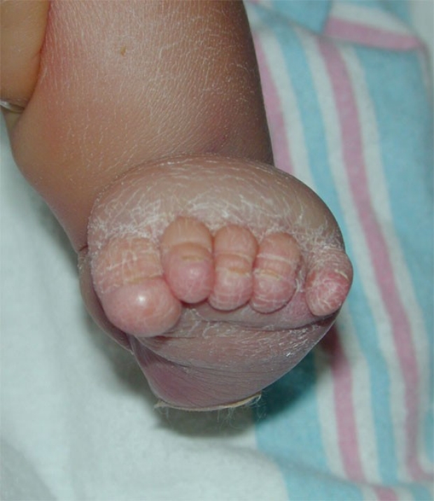
In this infant, the feet and lower legs seem unusually puffy. Although edema in general has a long list of possible etiologies, it is helpful to begin by determining if the edema is generalized or localized. Edematous hands and feet in particular are known to be associated with Turner's syndrome in infancy, so this diagnosis should be considered in girls with this finding. In this case, Turner's syndrome was the underlying etiology.
photo by David A Clark, MD
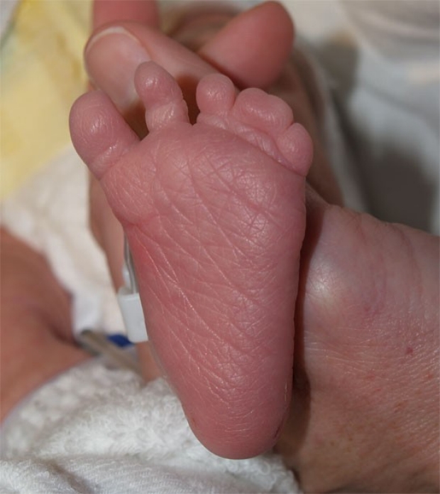
Here is another baby with edematous feet as a result of Turner's syndrome. From this angle, the excessive puffiness behind the toes can be easily seen.
photo by Division of Medical Genetics, Stanford University