Head
Head Circumference
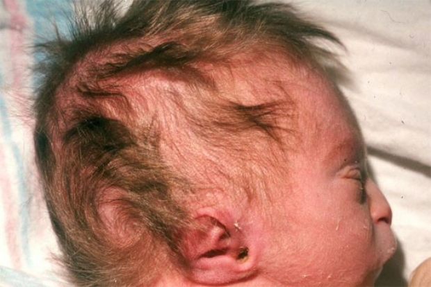
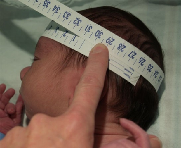
One of the first things to assess when evaluating the head of the newborn is the Occipital Frontal Circumference (OFC). This simple measurement may be the first clue to an underlying problem. The 50th percentile for OFC of a term newborn is 34 cm, so if an infant has a normal weight and length for a term infant (near 50th %ile for age), a measurement of <31 cm is disproportionately small (<< 10th %ile for age). Further evaluation is indicated; head imaging, screening for TORCH infection, and assessment for chromosomal abnormalities should all be considered.
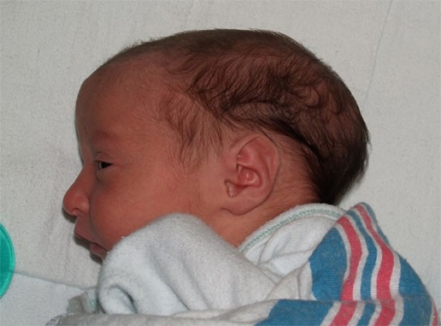
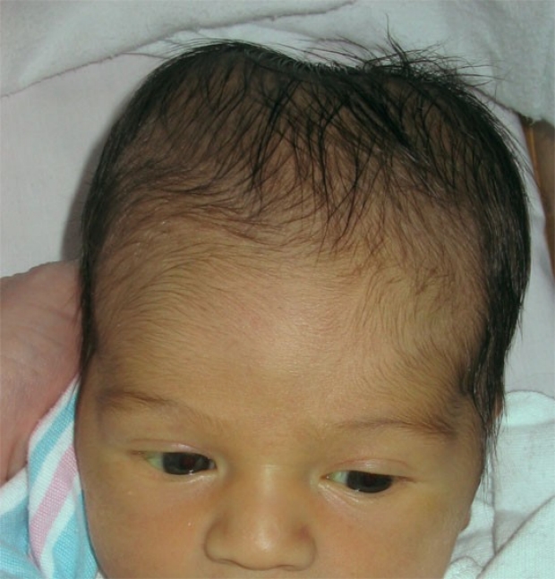
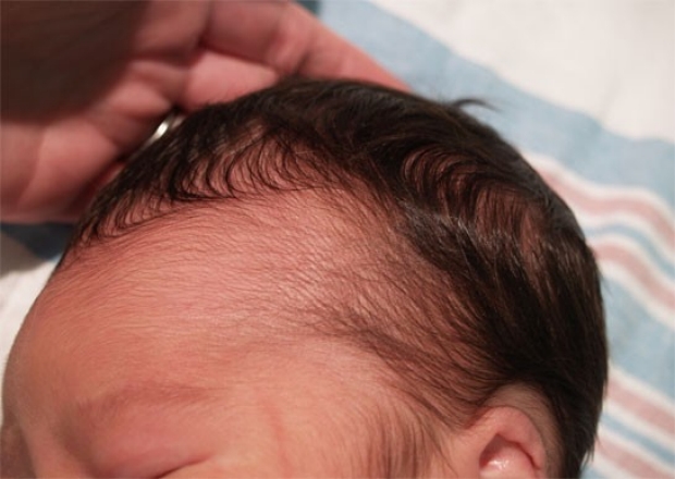
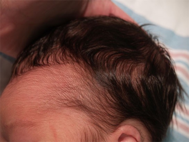
photo by Janelle Aby, MD