Genitourinary
-
Genitourinary
-
Normal Male Genitalia
-
Foreskin Variant
-
Megameatus
-
Hypospadias
-
Epispadias
-
Chordee
-
Webbed Penis
-
Buried Penis
-
Penile Torsion
-
Hypoplastic Urethra
-
Hydrocele
-
Inguinal Hernia
-
Acute Testicular Torsion
-
Testicular Torsion
-
In Utero Testicular Torsion
-
Descending Testicle
-
Ectopic Testicle
-
Undescended Testicles
-
Ambiguous Genitalia
-
Normal Female Genitalia
-
Vaginal Mass
-
Paraurethral Cyst
-
Prolapsed Ureterocele
This section contains pictures and/or video of newborn genitalia and is intended for educational purposes only.
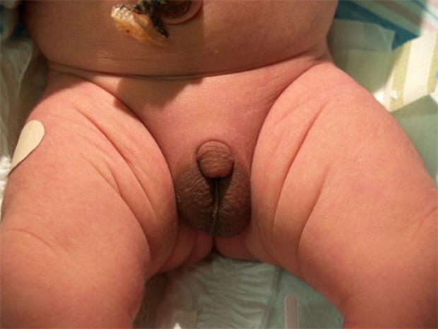
Normal Male Genitalia
Normal male genitalia in a term infant. The amount of pigment in the scrotum can vary considerably, depending on the ethnicity of the parents and maternal hormone effects.
photo by Janelle Aby, MD
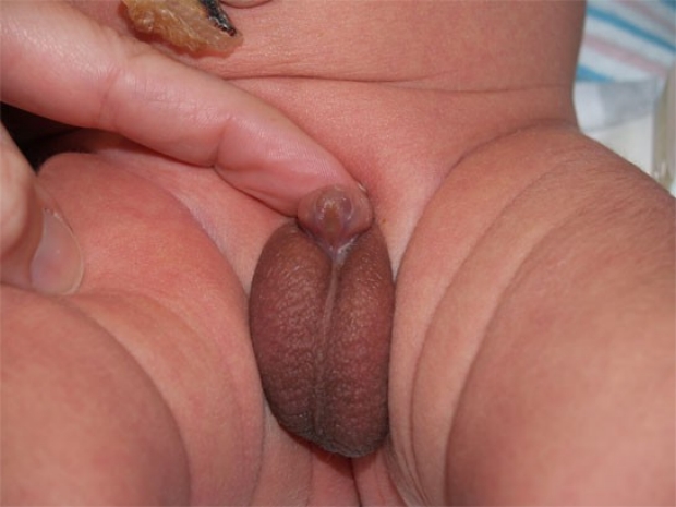
Foreskin Variant
Although the opening of the foreskin is usually tight enough in the newborn to preclude a view of the urethra, variablity exists. In this infant, a normal urethral meatus can be clearly seen through the wide opening of the foreskin. This is a variation of normal. No referral or intervention is necessary.
photo by Janelle Aby, MD

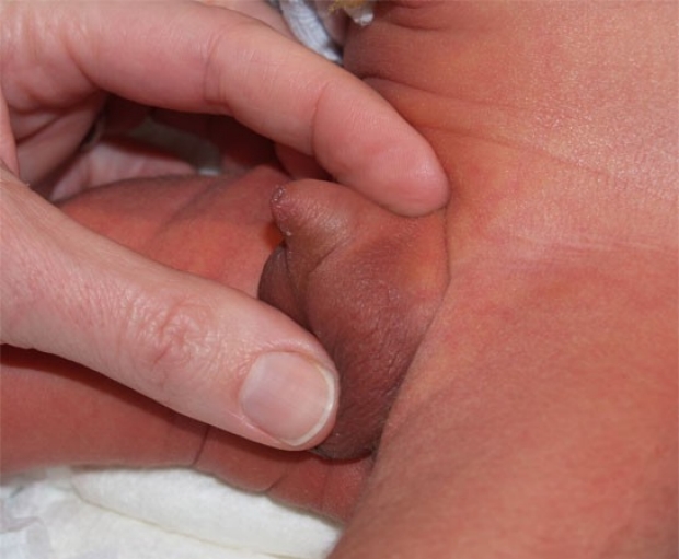
Megameatus
After retracting the skin for a circumcision, this anomaly of the meatus was noted. Although the meatus starts in a normal distal position, it extends down the ventral surface of the glans all the way to the corona. This is a variant of hypospadias, but it occurs with a normal foreskin.
photo by Janelle Aby, MD

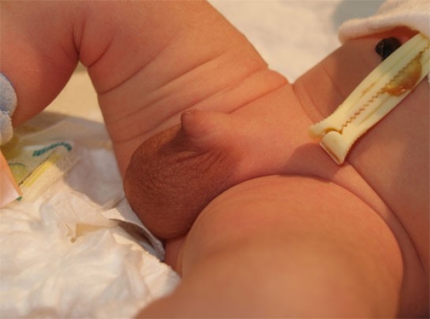
Megameatus
With use of a probe, the extent of the meatus can be more clearly seen. Circumcision is not contrindicated in this case, but the parents can be given the option of aborting the procedure in order to perform the circumcision at the time of the meatus repair or completing the circumcision as planned. There is no functional problem with this abnormality in the newborn period; repairs are usually planned when the infant is several months old, as the risk of general anesthesia is less at that age.
photo by Janelle Aby, MD
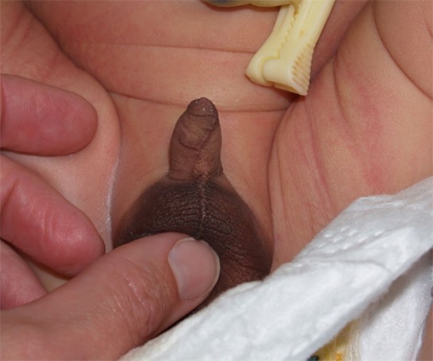
Hypospadias
Hypospadias with opening of the urethra on the distal penile shaft. Though not easily visible in this photograph, the urethral meatus is about midway between the normal location and the insertion into the scrotum. A hooded foreskin, which allows the glans to be readily visible, is present. The term "natural circumcision", though sometimes used, is a misnomer. The foreskin is present, just malformed. Hypospadias affects 1:250 boys, but most have a milder form than that shown here.
photo by LPCH housestaff
Hypospadias
This is another example of hypospadias. Here, the hooded foreskin can be readily appreciated. The urethral meatus is visible in a slightly more ventral position than is normal. This is a less severe degree of hypospadias, but still presents a contraindication to circumcision.
photo by David A Clark, MD
Hypospadias
In this patient, hypospadias is not readily apparent at first look, although the phallus does appear somewhat short for a full-term infant. The following photo demonstrates the abnormality more clearly.
photo by Janelle Aby, MD
Hypospadias
This is the same infant as in the previous photo. Here, slight pressure was applied on the dorsal edge of the base of the penile shaft to tip the phallus up. With this view, one can see that the ventral surface of the shaft seems to be largely absent and that the urethral opening appears to lie in the very short distance between the scrotum and the foreskin bulges on the sides of the glans.
photo by Janelle Aby, MD

Epispadias
With epispadias, the urethral meatus is dorsally displaced. This condition is much less common than hypospadias, but also presents a contraindication to routine circumcision. In it's severe forms, epispadias is often associated with exstrophy of the bladder.
photo by William Kennedy, MD

Epispadias
This is an example of severe epispadias and bladder exstrophy. The penis appears to be nearly completely splayed open lengthwise on the dorsal surface, and actually has a dorsal curvature which is minimized by the downward pressure applied to the scrotum. The red tissue inferior to the umbilical cord is the inner (posterior) wall of the bladder which is also splayed apart and protruding through a defect in the anterior pelvic wall. Surgical correction is required.
photo by William Kennedy, MD

Chordee
Chordee exists when there is a ventral curvature of the penis. It can occur in the setting of hypospadias, but can also be isolated, as in this case. In this baby, the curvature is most visible from this view from the side. Chordee is a contraindication to circumcision in the newborn period, but if desired, circumcision can be done at the time of surgical correction.
photo by Janelle Aby,MD
Chordee
This is a frontal view of the same infant. The tip of the foreskin is not easily appreciated in the photograph, but on examination the foreskin and meatus appeared to be normal. Curvature of the shaft is the only abnormality in this case.
photo by Janelle Aby,MD

Webbed Penis
When present, a penoscrotal web (webbed penis) is another contraindication to routine circumcision in the nursery. On cursory examination, the penis in this child appears to be within normal limits; however, there was very little skin surface between the tip of the foreskin and the scrotum. The following photo shows the abnormality more clearly.
photo by Janelle Aby,MD
Webbed Penis
When traction is applied to the skin at the base of the penis, the web can more clearly be seen. Routine circumcision is contraindicated in this case as it may lead to the penis becoming trapped behind a scarred, tent-like fold of skin.
photo by Janelle Aby,MD

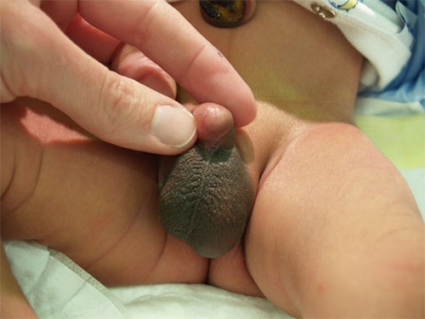
Buried Penis
In some infants, the penis appears to be "buried" in the tissues surrounding it. This is another contraindication to routine circumcision, as it could result in the penis becoming trapped underneath the healing circumcision site and result in an overall worsening of the condition.
photo by Janelle Aby,MD
Buried Penis
Here is another example of a newborn with a buried (concealed) penis. With this view, the ventral foreskin looks unusually short and the shaft of the penis is not readily visible.
photo by Janelle Aby,MD

Buried Penis
When downward pressure is applied beside the penile shaft, more of the underlying normal anatomy can be seen. However, because of the tendency of the penis to retract back into its previous position and because of the relatively small amount of foreskin present, this is still a contraindication to routine neonatal circumcision.
photo by Janelle Aby,MD

Penile Torsion
This is an example of penile torsion of about 90 degrees. Although the urethral meatus itself cannot be visualized, one can see that the relatively flat ventral surface of the glans is facing the infant's left thigh instead of the midline. A mild degree of torsion (<60 degrees) is felt to be a variation of normal and is not a contraindication for circumcision. In this case, circumcision was postponed until the time of surgical correction at about 4 months of age. Penile torsion does not cause functional impairment in newborns, but high degrees of torsion should be corrected (with or without circumcision) to prevent painful or dysfunctional erections in later life.
photo by Janelle Aby,MD
Penile Torsion
Another clue to a possible penile torsion is the rotated appearance of the median raphe. Although the raphe frequntly zigzags a bit, it usually starts and ends at or near the midline. Here, the raphe starts in the midline at the scrotum, but then winds around the shaft to the left and ends up on the left side at tip. Though not a diagnosic finding, this appearance should at least lead the examiner to evaluate more closely.
photo by Janelle Aby,MD
Penile Torsion
Because circumcision was desired and penile torsion was suspected, a urology consult was obtained before this procedure. All agreed that the likely torsion was about 45 degrees and that circumcision could safely be done in the nursery. Here the counterclockwise rotation of the urethral meatus can be seen in relation to the midline of the scrotum.
photo by Janelle Aby,MD
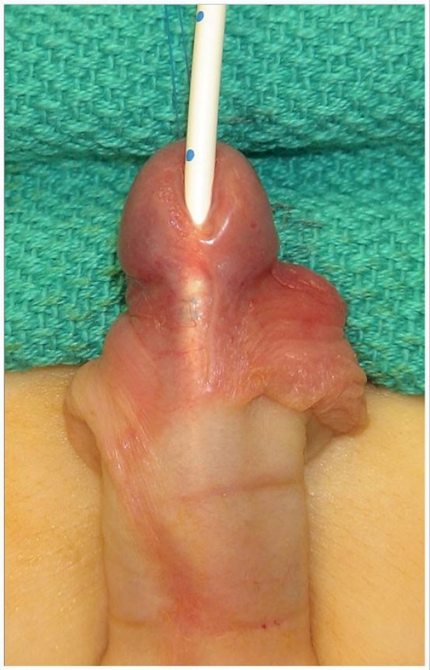
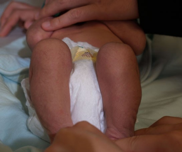
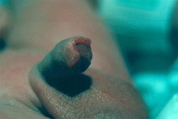
Hypoplastic Urethra
When examining the penis, assessment of the adequacy of skin circumferentially should be made. If the ventral side seems deficient, urethral hypoplasia may exist. Patients with this condition often have a penile raphe that zigzags back and forth as well as a thinned or shortened foreskin on the ventral side. If in doubt, a small feeding tube can be threaded into the urethra. If the print on the tube is visible through the skin, as it is in the photo above, circumcision is absolutely contraindicated, and a pediatric urologist should be consulted.
photo by William Kennedy, MD

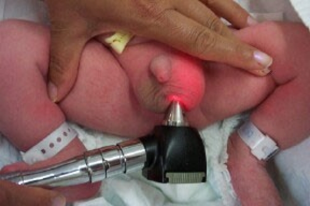
Hydrocele
Hydroceles are a frequent finding in newborns. It is important on palpation to identify the normally small testicles (approximately 1 cm) as separate entities from the large, smooth-walled fluid collections of hydroceles. In contrast to inguinal hernias, common (non-communicating) hydroceles cannot be reduced as the fluid is in an enclosed space. Spontaneous resolution is expected.
photo by Janelle Aby,MD

Hydrocele
Here one can see how easily the fluid-filled hydroceles transilluminate. Transillumination may be used to assist in making a diagnosis but for an experienced examiner, the diagnosis is usually made by palpation alone.
photo by Janelle Aby,MD
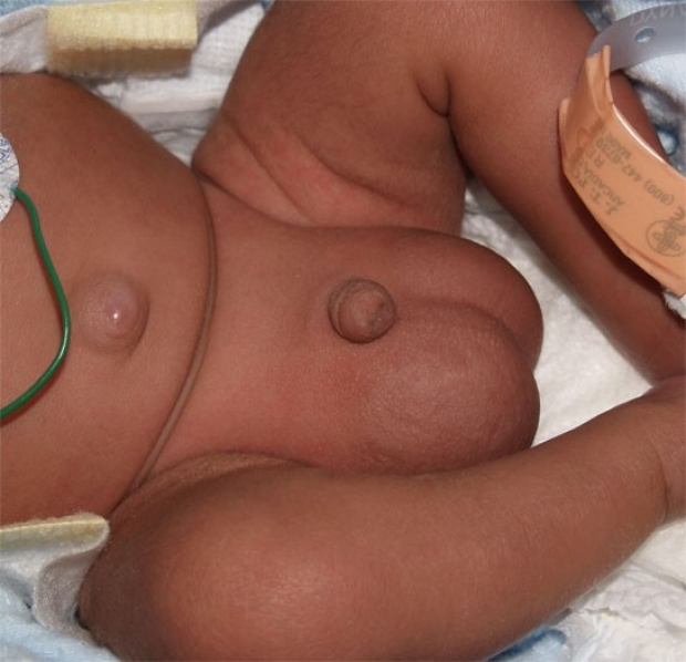
Inguinal Hernia
This is a typical appearance of bilateral inguinal hernias. In contrast to hydrocele, inguinal hernias are typically associated with a fullness in the inguinal area. This infant is preterm, a risk factor for the presence of hernias. While the scrotum may transilluminate with hernias, the masses can frequently be reduced with digital pressure. At times bowel sounds may also be appreciated over the area of the mass.
photo by Janelle Aby,MD

Acute Testicular Torsion
This otherwise well infant presented with acute color change of the scrotum. The right testicle was torsed and the baby was taken emergently to surgery. Surprisingly, infants with acute torsion may not exhibit signs associated with pain; as in this case, this condition may be an unexpected finding on routine physical examination.
photo by Anthoy Burgos, MD
Acute Testicular Torsion
The same infant as in the previous photo. Because of the pathology that is occuring in the right testicle, the right scrotum does not transilluminate equally with the left.
photo by Anthony Burgos, MD

Testicular Torsion
This is another example of testicular torsion in the newborn. Here, the discoloration is more subtle, but the difference in size between the two halves of the scrotum is striking. The testicle on the baby's right side is the affected one.
photo by David Clark, MD
In Utero Testicular Torsion
With antenatal torsion, the scrotum typically has a normal appearance. On examination, however, the right testicle was slightly larger and more mass-like in texture than the normal left testicle. Ultrasound with doppler confirmed that there was no blood flow to the right side. This infant was taken to the OR on day 2 for removal of the right testicle and fixation of the left testicle as the contralateral side is also at increased risk of torsion.
photo by Janelle Aby, MD

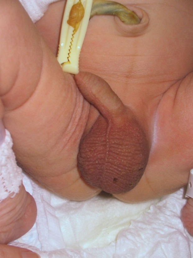
Descending Testicle
In a term infant, the testicles have normally completed their migration from the abdomen into the scrotum. However, even term infants will at times have one testicle that is undescended or incompletely descended. In this infant, the right side of the scrotum was empty and the testicle was palpated in the inguinal canal (seen as a bulge in the photo above). Because this testicle is located along the normal pathway of descent and is not ectopic, no further evaluation is needed. Spontaneous descent into the scrotum is expected with time. Circumcision is permissible in this situation.
photo by Janelle Aby, MD
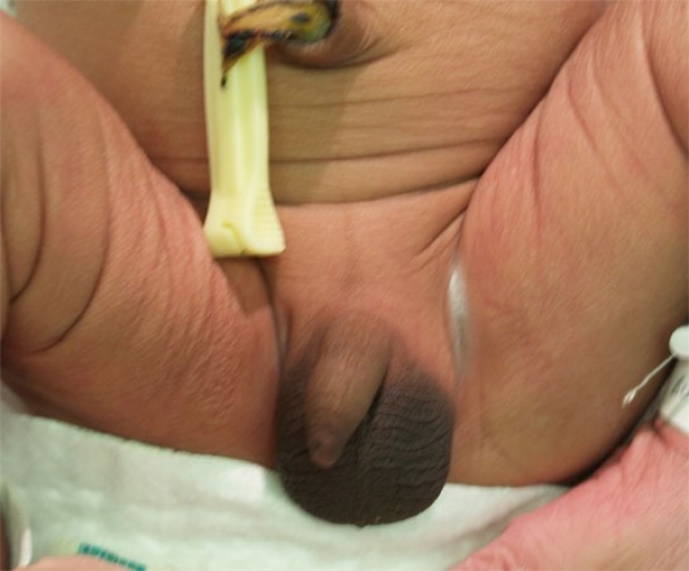
Ectopic Testicle
During descent, it is possible for the testicle to pass through the inguinal ring and then veer off course into an aberrent location. On examination of this infant, the right testicle was absent from the scrotum, but a mass was appreciated over the perineum. Ultrasound confirmed the diagnosis of ectopic testicle. Ectopic testicles may be found in the superficial inguinal pouch, the suprapubic area, the perineum, the femoral canal, or even the other side of the scrotum. This may eventually require surgical intervention, but it does not present a contraindication to neonatal circumcision if the parents desire it. (Note the fresh circumcision site in the photo!)
photo by Janelle Aby, MD

Undescended Testicles
At times, a testicle is not palpable at all in the scrotum. In the photo above, the scrotum appears asymmetric with the right side flatter than the left. Palpation confirmed that a testicle was present on the left, but not on the right. Unilateral undescended testicle is fairly frequent finding in term newborns and does not require further evaluation at birth or preclude circumcision. With time (weeks to several months) the testicle is expected to descend spontaneously. Bilateral undescended testicles, on the other hand, are quite unusual at term and should be considered as ambiguous genitalia (with circumcision contraindicated) until proven otherwise.
photo by Janelle Aby, MD
Ambiguous Genitalia
In any case where the genitalia are ambiguous, a work-up to determine gender and the underlying cause of the ambiguity should begin immediately. These infants should be initally observed in the NICU so that any electrolyte abnormalities can be identified and treated rapidly, and appropriate specialists involved (e.g. endocrine, genetics, urology). Circumcision is absolutely contraindicated when genital ambiguity exists. This particular infant was an under-virilized male with hypospadias, micropenis, and bifid scrotum (testes were present bilaterally).
photo by Janelle Aby, MD

Ambiguous Genitalia
This baby has genitalia that appear similiar to the previous photo, but this infant was a virilized female with 21 hydroxylase deficiency.
photo by David A. Clark, MD
Ambiguous Genitalia
This infant is a male with micropenis and a cleft scrotum, but on initial evaluation the gender cannot readily be determined. Genetic, metabolic, and urologic evaluation is necessary.
photo by David A. Clark, MD
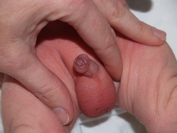
Normal Female Genitalia
Normal female genitalia in a term infant. Some vernix is visible in the right inguinal crease.
photo by Janelle Aby, MD

Normal Female Genitalia
The increased pigment over the labia in this African-American newborn is normal. Both the color of the parents and exposure to maternal hormones in utero impact the appearance, so a wide variation in pigment tones is expected. Those babies with more melanin tend to have increased pigment in several places: helices of the ears, bases of the nails, in the axillae, at the edge of the umbilical stump, and over the scrotum or labia. Maternal hormone exposure produces the linea nigra (down the middle of the abdomen) that can also be seen in this picture.
photo by Janelle Aby, MD
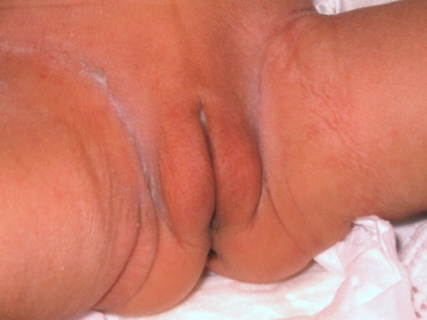
Normal Female Genitalia
When examining the genitalia of a girl, it is important to visualize the hymenal tissue and labia minora. The hymenal tissue is the light pink tissue that can be seen between the labia minora. It should appear to have a central opening, as it does here. White vaginal discharge (as seen around the hymen here) is extremely common in the newborn period. This infant also has some vernix (a cheesy white material) on the interior surface of the labia majora. No special cleaning is required.
photo by Janelle Aby, MD

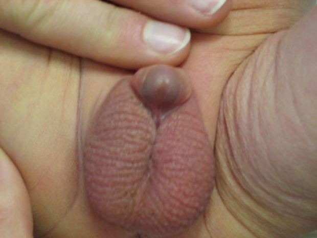
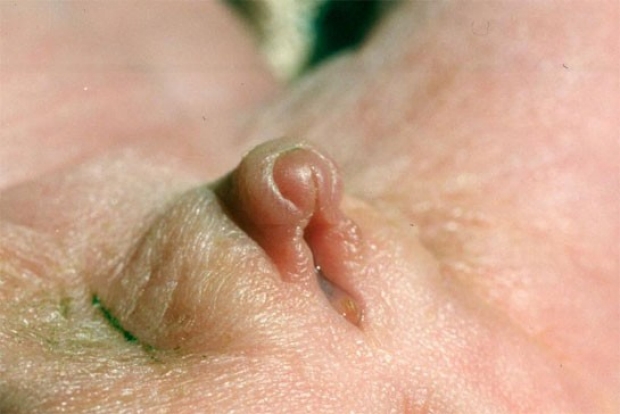
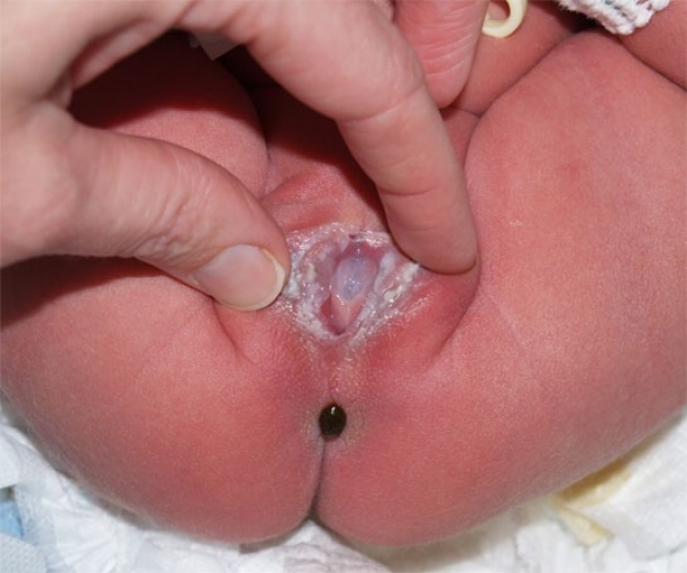
Vaginal Mass
This mass was noted on the discharge examination in the newborn nursery. It was not readily visible on casual examination of the external genital, but with increased abdominal pressure from crying or with closer inspection of the genitalia, it could be seen. Voiding was not impaired. The imaging studies in this case gave conflicting results, but it most likely represented a multiloculated cyst. The small, light pink tissue inferior to the mass is a hymenal tag, a normal finding.
photo by Janelle Aby, MD
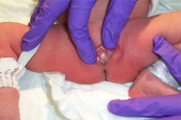
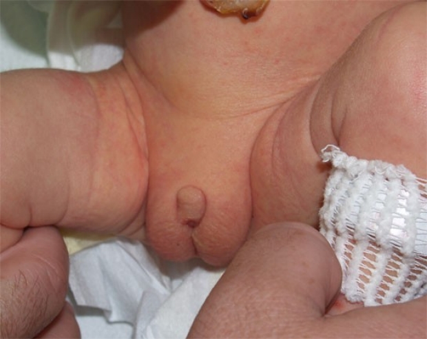
Paraurethral Cyst
This mass, present at birth, was a paraurethral cyst. The fact that the hymen can still be seen inferior to the mass helps differentiate this from imperforate hymen, which could have a similar appearance.
photo by Priya Akula, MD

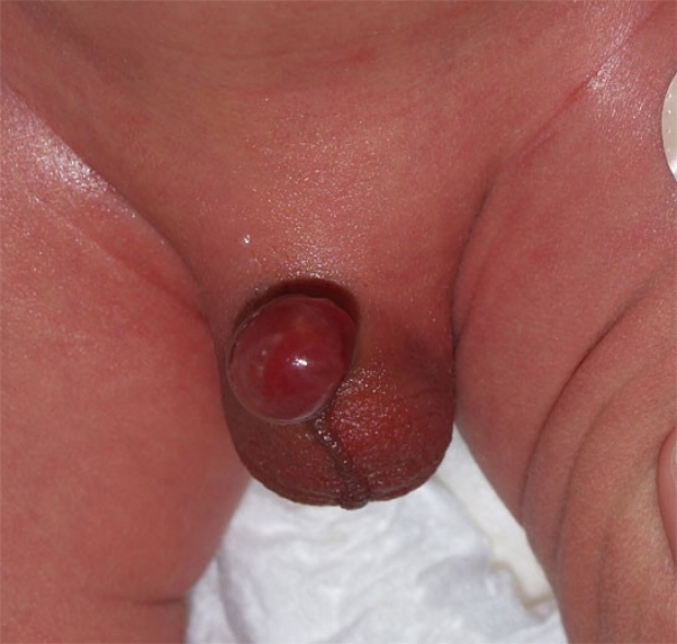
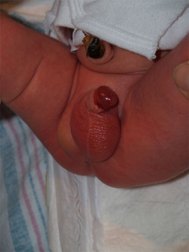
Prolapsed Ureterocele
This mass is large enough to obscure all intra-labial landmarks, and is quite hemorrhagic in appearance. This is a ureterocele which has actually prolapsed through the urethra to be visible externally. Surgical intervention is necessary in this case.
photo by William Kennedy, MD