Researchers at Stanford demonstrated that direct stimulation of fewer than two dozen neurons linked to social interaction was enough to suppress a mouse’s drive to feed itself.
January 16, 2019 - By Bruce Goldman
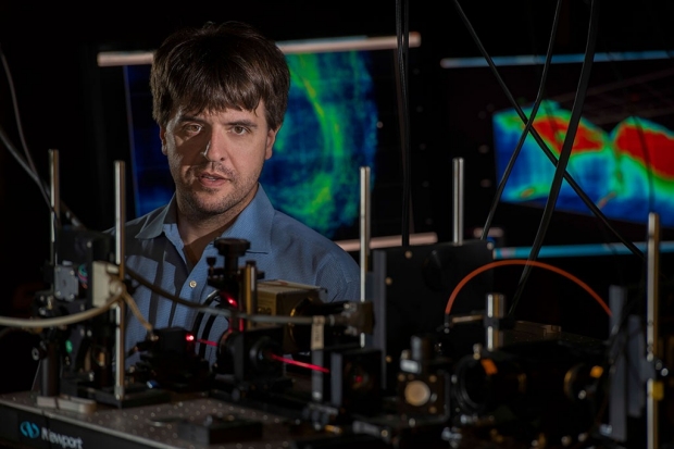
Karl Deisseroth and his team have used a method for stimulating and monitoring several neurons at once to identify different clusters of neurons responsible for feeding behavior and social behavior.
Steve Fisch
Feeding behavior and social stimulation activate intermingled but distinct brain circuits, and activating one circuit can inhibit the other, according to a new study by researchers at Stanford University.
The researchers demonstrated in mice that direct stimulation of fewer than two dozen nerve cells, or neurons, linked to social interaction was enough to suppress the animals’ drive to feed themselves — a finding with potential clinical significance for understanding and treating eating disorders such as anorexia.
The researchers made these findings by developing a technique for teasing apart separate but closely intertwined sets of neurons in the brain.
A paper detailing the findings and the method used to obtain them was published online Jan. 16 in Nature. The senior author is Karl Deisseroth, MD, PhD, the D.H. Chen Professor and professor of bioengineering and of psychiatry and behavioral sciences and a Howard Hughes Medical Institute investigator. Lead authorship is shared by postdoctoral scholars Joshua Jennings, PhD, and Christina Kim, PhD, along with staff scientist James Marshel, PhD.
Social curbs on eating behavior
“We know social situations can inhibit the urge to eat,” Deisseroth said. “One example is the behavior of people at different levels of dominance in a social hierarchy. You’re not going to dive into that plate of ribs when you’re dining in the presence of royalty.”
Anorexia is another example. “People with anorexia report that a powerful driver, at the disorder’s onset, was feedback from others indicating they’d be rewarded for restricting their food intake,” said Deisseroth.
Virtually nothing is known about the neural underpinnings of this inhibition, he said. “We sought to understand, at the level of individual neurons, how these potentially competing drives may negotiate with each other, and how the brain circuits associated with feeding versus social behavior may interact.”
A key goal was to know which neurons actually matter for behavior.
Deisseroth’s group focused on a part of the brain called the orbitofrontal cortex, a sheet of cells that, in both mice and humans, lies on the brain’s outer surface toward the front of the organ. This brain region, which is similar in the two species, has been shown in human imaging studies to be active when subjects are wishing for, seeking, obtaining and consuming food, or when they’re socially engaged.
Exploring the interactions of feeding and social drives was guaranteed to be tricky.
“It’s not as if there is a cluster of ‘feeding’ neurons’ and another cluster of ‘social’ neurons sitting in two neatly labeled clumps in the orbitofrontal cortex, so you can just position an electrode in one or the other cluster and find out all you need to know,” Deisseroth said. The neurons driving and responding to these different activities are interspersed, scarce and scattered throughout the orbitofrontal cortex like sprinkles on a cupcake. Plus, they all look pretty much the same.
So the researchers designed a sophisticated system for simultaneously stimulating and monitoring activity in multiple designated neurons. This let them determine which orbitofrontal-cortex neurons were active during feeding-associated or social activities, or both, or neither. The technology also allowed them to stimulate on the order of 20 neurons identified as dedicated to one or the other activity and watch what behavior resulted.
Over the past decade and a half, Deisseroth has pioneered the development of an experimental approach called optogenetics, in which a gene for a light-sensitive protein called an opsin is inserted into neurons so they can be activated by pulses of laser light reaching them via an implanted optical fiber. Recent advances in his lab have optimized one such opsin to the point where his team can stimulate numerous selected, behaviorally categorized neurons at a time in a mammal.
“This study builds on our initial demonstration in mammals of single cell control with optogenetics in 2012, but now marks the first demonstration of control of mammalian behavior by the manipulation of multiple, individually specified neurons,” he said.
The scientists inserted the gene for this improved opsin into the orbitofrontal cortex of mice, along with another gene that causes neurons to fluoresce in proportion to their activity. A tiny lens at the tip of the optical fiber guided light across numerous targeted neurons with near simultaneity, causing as many as two dozen designated neurons to fire together.
Food vs. friends
During the experiments that followed, the mice were constrained by an apparatus that kept their heads comfortably fixed in place. In one set of experiments, mice were exposed to a spout that occasionally issued a drop of a high-calorie solution, which could be readily licked up. For each mouse, Deisseroth’s colleagues recorded which orbitofrontal-cortex neurons among the several hundred in their field of view lit up during this activity.
Optogenetically stimulating just 20 feeding-responsive neurons enhanced the mice’s licking activity in the presence of the high-calorie solution, tying those neurons causally to feeding behavior.
To identify social-responsive neurons in the mice’s orbitofrontal cortex, the scientists introduced juvenile mice — which older mice perceive as nonthreatening potential buddies and set about sniffing — and tracked activity levels in the neurons in the field of view. They were able to identify specific neurons responsive to the exploratory social interaction.
Optogenetically stimulating social-responsive orbitofrontal neurons in the presence of a caloric reward reduced the amount of time the mice spent licking the solution. So did the natural-stimulation equivalent: exposure to juvenile mice. The more the social interaction, the less the interest in calories.
While the mice in this study weren’t a disease model, Deisseroth noted the findings’ potential clinical significance.
“We’ve been able to pinpoint otherwise indistinguishable orbitofrontal-cortex neurons involved in feeding and social drive states,” he said. “A key goal was to know which neurons actually matter for behavior. Now that we do, we can examine them more closely to look for, say, surface-protein markers or wiring differences that distinguish them from one another. If there are any distinctions like that, it will deepen our understanding of how competing drives are negotiated among neuronal cell types in the cerebral cortex — and could even lead to pharmaceutical interventions that reduce social inhibition of food consumption among people with anorexia.”
Deisseroth is a member of Stanford’s Wu Tsai Neurosciences Institute and of Stanford Bio-X.
Other study co-authors are graduate student Misha Raffiee; postdoctoral scholar Li Ye, PhD; senior computational and optical systems engineer Sean Quirin, PhD; life science research professional Sally Pak; and life science research assistant Charu Ramakrishnan.
The study was funded by the National Institutes of Health (grants R37DA035377, R37MH075957 and R01MH086373), HHMI, the Defense Advanced Research Projects Agency, the National Science Foundation, the Wiegers Family Fund, the Nancy and James Grosfeld Foundation, the Sam and Betsy Reeves Fund and the H.L. Snyder Foundation.
Stanford’s departments of Bioengineering and of Psychiatry and Behavioral Sciences also supported the work. The Department of Bioengineering is jointly managed by the School of Medicine and School of Engineering.
-
 Bruce GoldmanBruce Goldman is a senior science writer in the Office of Communications. Email him at goldmanb@stanford.edu.
About Stanford Medicine
Stanford Medicine is an integrated academic health system comprising the Stanford School of Medicine and adult and pediatric health care delivery systems. Together, they harness the full potential of biomedicine through collaborative research, education and clinical care for patients. For more information, please visit med.stanford.edu.

