Researchers engineered stem cell-derived heart tissues to study how tachycardia affects the heart and to uncover the inner workings of our body’s engine.
December 19, 2023 - By Andrea Tamayo
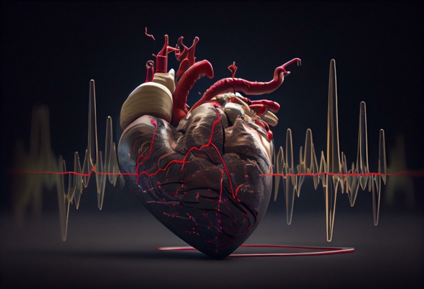
To study tachycardia-induced cardiomyopathy, Stanford Medicine researchers engineered heart cells from human stem cells.
Caleb
Heart rates are easier to monitor today than ever before. Thanks to smartwatches that can sense a pulse, all it takes is a quick flip of the wrist to check your heart. But monitoring the cells responsible for heart rate is much more challenging — and it’s encouraged researchers to invent new ways to analyze them.
Joseph Wu, MD, PhD, director of the Stanford Cardiovascular Institute and professor of medicine and of radiology, has devised a new stem cell-derived model of heart tissue that provides insight into conditions that crop up when heart cells beat out of control. In particular, Wu is studying a disorder called tachycardia, which increases the heart rate and can lead to cardiomyopathy, in which the heart loses its ability to pump blood sufficiently in people with otherwise healthy heart structures.
“Tachycardia is probably more common than we think,” said postdoctoral scholar Chengyi Tu, PhD, who helped lead the work. “It’s believed to be underdiagnosed because an increase in heart rate is quite common in different types of heart diseases, and it gets masked.”
To study tachycardia-induced cardiomyopathy, the researchers engineered heart cells from human stem cells to uncover how our body’s engine runs when it’s in overdrive.
Joseph Wu
“Modeling tachycardia-induced cardiomyopathy with human stem cell-derived heart tissues allows us to better understand the impact of fast heart rates on our bodies,” said Wu, the Simon H. Stertzer, MD, Professor who is the senior author of the study. It was published Nov. 27 in Nature Biomedical Engineering. Tu is the lead author.
Engineering heart cells
Unlike most types of organ tissues, heart cells are extremely difficult to grow in a lab. Patient heart cells cultured in a dish tend to de-differentiate –– or lose their primary function and fail to beat.
“Ideally, you want to take samples of a patient’s heart just after disease diagnosis, during illness and after treatment,” Tu said. “To validate your discovery, you need a lot of replicates to give you statistical power, but clinically, it’s impossible to sample so frequently.”
Given the dearth of tissue, Wu and his colleagues grew more than 400 heart tissue samples from stem cells to look at how heart cells function, a process that spanned more than four years.
“Making engineered heart tissue is very different from culturing cells in a dish. The timeline is very long,” Tu said. Generating heart cells from stem cells takes about two weeks; putting them together in a 3D tissue and maturing them takes almost two months.
Restoring the chemical balance
Using a wired chamber, the researchers electrically stimulated the cells, inducing tachycardia. They tested whether the cells could recover from tachycardia over the course of 10 days. During the first five days, the cells’ ability to contract continuously declined to about 50% of normal function. But once the researchers stopped the electrical stimulation, the cells made a full recovery in five days.

Chengyi Tu
That tracks with what doctors already know about tachycardia-induced cardiomyopathy — it’s mostly reversible. When a person’s heart rate slows back down, their heart tissue function returns to normal.
In another experiment, researchers induced tachycardia in a different group of engineered heart tissue. Then, after stopping stimulation, the team supplemented the tissues with NAD –– a molecule that supports energy reactions –– and saw the heart cells’ function recover more rapidly. The supplemented tissues had recovered 83% of their original function by the first day, while the untreated group showed little improvement.
To validate their findings, the team compared the engineered heart tissues with clinical human data and canine model data. “I was surprised by how well the engineered heart tissues mimic the real human hearts,” Tu said.
Uncovering the molecular switch
During tachycardia, the heart may struggle to pump blood to the rest of the body because the fast heart rate prevents the heart’s chambers from filling up and contracting fully. If it persists for several days or weeks, which can happen in severe cases, blood vessels stop supplying enough oxygen to the heart tissue and the rest of the body.
When beating normally, the heart uses fat as an energy source, but breaking down fats requires a lot of oxygen. Without oxygen, the heart’s fuel source switches to sugar in a process called metabolic rewiring. The fuel switch and hypoxia, or lack of oxygen, contribute to a decrease in the NAD/NADH ratio, a vital chemical duo that helps maintain the function of a protein in heart tissue known as SERCA.
“Varying levels of the SERCA protein act like a gas and brake pedal for a car,” Tu said. When researchers increase the amount of NAD, the heart’s gas pedal is thrusted, and the SERCA protein strengthens the heartbeat of the engineered cells. When decreased, the engineered heart tissues hit the brakes, making them beat more weakly.
By giving patients NAD through an off-the-shelf supplement or by IV injection, clinicians believe they can restore the chemical balance and accelerate a patient’s recovery.
Alongside a new possible supplement to help patients recover from tachycardia, the research demonstrates the importance of new methods to model disease. Last year, President Joe Biden signed the FDA Modernization Act 2.0 into law, which removed the requirement for animal testing before human drug trials. “Now, there is more need for non-animal models to complement the animal models,” Tu said. “This work proves that it’s possible to model complex cardiac conditions using a universal non-animal model to study this disease and test possible therapeutics.”
This study was funded by the American Heart Association, the National Institutes of Health (grants K99 HL164962, K01 HL130608, R01 HL151345, R01 HL163680, R01 HL141371, R01 HL113006, R01 HL150693 and P01 HL141084), and the National Aeronautics and Space Administration.
About Stanford Medicine
Stanford Medicine is an integrated academic health system comprising the Stanford School of Medicine and adult and pediatric health care delivery systems. Together, they harness the full potential of biomedicine through collaborative research, education and clinical care for patients. For more information, please visit med.stanford.edu.

