Radiological Sciences Laboratory Internship Projects
Summer 2024 Research Experience for Undergraduates (REU) EXTENDED DEADLINE March 29, 2024
Program Requirements:
This is a 10-week program starting June 24 and ending August 30, 2024 (40 hours per week). Students are expected to participate for the duration of the program (exceptions may be granted at the discretion of the faculty mentor). Students are also required to present their research at the final presentation/poster session and complete a program questionnaire.
Selected students will receive a $7500 stipend for the 10-week (40 hours per week) summer session of full-time research work. Stipend funding is provided by the Stanford VPUE office and from the RSL division. All funds are managed through the Stanford Financial Aid Office.
Who can apply?
Stanford Undergrads who will not have graduated by the beginning of the program. Co-term students may also apply. Stanford undergrads must have been enrolled full-time for a minimum of 2 quarters in the 2023-2024 academic year. Note: students on a Leave of Absence (LOA) or who have been on a LOA for 3 consecutive quarters prior to the funding period are not eligible to receive funding. Housing is not provided (Students may enroll separately for Stanford -based housing).
Undergraduate students from other institutions who have 2 years of college experience (i.e. rising juniors or seniors) and have not graduated by the beginning of the program. International students who are matriculated at U.S. Colleges or U.S. Universities may apply (visa restrictions may apply).
How to apply:
- Review the open projects below. You may apply for 1-3 projects.
- Create a letter of interest including your full name, college or university, academic year, and area of study (or interest). Also, describe your interest in this project in a short paragraph. Be sure to include: what aptitude and skills you have for the project and how the REU experience correlates with your current and future academic ambitions.
- Send your letter via email to: bbonini@stanford.edu. The subject line must include the LAB IDENTIFIER listed with each project. HINT: letters are best sent individually as one project per email.
- Applications: Round 2 now open through March 29, 2024
- All inqueries will be reviewed and qualified students will be invited to apply. The application process will request a transcript, resume, classwork completed, references, and demographic information. Preview the application HERE
- Qualified applicants will be contacted directly by the sponsoring faculty. Acceptance notices will be sent on a rolling basis. All applicants will receive a response.
Program Highlights
RSL is offering the following enrichment opportunities to our 2024 Summer Interns.
Students will be given time away from lab work:
- to learn about diverse imaging modalities through the Introductory Lecture Series
- to attend RSL Group Meetings and selected lectures.
- to develop scientific presentation skills and more at specialized mentoring sessions.
- to attend graduate school admissions and information sessions
- to have weekly lunches with RSL professors.
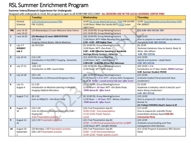
Example Schedule
Questions?
Please direct any questions concerning the REU Program, including the application process to bbonini@stanford.edu
| LAB GROUP | PROJECT STATUS | NEW or REVISED | |
| Airan | Open | in review | |
| Boada | Open | in review | |
| Butts Pauly | Open | 03.01.24 | |
| Dahl | Open | 03.01.24 | |
| Daniel/Hargreaves | Open | 03.01.24 | |
| Ennis | Open | 03.01.24 | |
| Glover | TBA | 03.01.24 | |
| Gold | Open | 03.01.24 | |
| Kogan | Open | 03.01.24 | |
| McNab | Open | 03.01.24 | |
| Moseley/Zaharchuk | Open | 03.01.24 | |
| Rutt | Open | 03.01.24 | |
| Setsompop | Open | 03.01.24 | |
| Spielman | Open | in review | |
| Wang | Open | 03.01.24 | |
| Zeineh | Open | 03.01.24 |
Faculty Mentor: Adam Wang, PhD
Internship Mentor: Adam Wang, PhD; TBN
IDENTIFIER: WangLab
Type of Laboratory Research: Dry lab
The Wang group develops technologies for advanced x-ray and CT imaging, including artificial intelligence (AI) methods, novel system designs, and spectral CT imaging. We develop these technologies with advanced computational tools, in the Photon Counting CT Lab (a next-generation prototype CT scanner for human subjects and phantoms), Zeego Lab (a robotic C-arm that supports interventional imaging, animal studies, and more), and in the Tabletop X-Ray CT Lab (for prototyping novel systems, custom geometries, and phantom experiments).
The REU project will be customized to the student’s skills and interests, and a lab mentor will be identified accordingly. Possible computational projects include using AI to improve CT image quality, reconstructing images from novel CT systems, or optimizing CT for radiation dose and image quality. Possible lab projects include developing open-source x-ray technologies, 3D printing of realistic anatomy, or evaluating spectral CT detectors.
Required skills:
Seeking highly motivated students enthusiastic about research. Suitable for students with EE, BioE, Physics, CS, or similar backgrounds. Familiarity with Python, Matlab, or C++ programming languages.
Faculty Mentor: Fernando E. Boada, PhD
Internship Mentor(s): Fernando E. Boada, PhD, TBD
IDENTIFIER: BoadaLab
Type of laboratory research: Image Reconstruction Algorithm Development (Dry Lab).
Project Title: Anatomically-Guided Image Reconstruction Project Description: The goal of the project is to develop the first prototype of an artificial intelligence (AI) application to provide improved resolution and sensitivity for physiologically-based brain scans (e.g., sodium MRI, Amyloid PET scans) using the information from high-resolution MRI scans as a constraint. Specific, short-term, applications include brain tumors, Alzheimer’s disease and Stroke.
Overview of lab work: Our lab focuses on the development of image acquisition and reconstruction technologies for providing physiological information for the brain that cannot currently be provided using state-of-the-art conventional MRI and/or PET imaging techniques. Our lab has a strong focus on Neuropathology (e.g., Stroke, Brain Tumors, Sodium-ion Imbalances); in this setting the use of advanced image reconstruction techniques such as Anatomically-Guided Reconstructions (AGR) enables techniques such as sodium MRI to be used within the confines of a clinical environment to answer questions about pathophysiology that are not properly addressed using conventional MRI techniques (e.g., tumor proliferation). Projects are developed using the Anaconda (Python) environment, which enables efficient algorithm development and testing. There will be constant and close collaboration with clinicians to facilitate development and evaluation of the image reconstruction tools.
Required Skills: Willingness to work in a multi-disciplinary team, curiosity, altruism, friendliness, integrity and self-motivation are essential. Basic familiarity with computers and Python is essential.
_________________________________________________
Faculty Mentor: Daniel Spielman, PhD
Internship Mentor: Daniel Spielman, TBN
IDENTIFIER: SpielmanLab
Title/Project: Magnetic resonance methods for measuring brain energy metabolism and oxidative stress.
Beyond anatomy, magnetic resonance imaging (MRI) and spectroscopy (MRS) provide unique opportunities for assessing metabolism within the body. You will be joining a multidisciplinary research team with expertise spanning physics, electrical engineering, biochemistry, and clinical sciences, to help develop novel data acquisition and processing tools for assessing critical in vivo processes. Our lab’s primary focus is on imaging in vivo metabolism throughout the body. Projects include applying machine learning for the improved quantitation of MRS, multinuclear MRI of brain energy metabolism in brain tumors, and understanding brain metabolism during pediatric cardio bypass surgery. Primary applications of this work include 1) improved understanding and treatment of neurologic conditions including traumatic brain injury, Alzheimer's disease, schizophrenia, and autism, 2) advanced brain tumor diagnosis and the assessment of response to therapy, and 3) minimizing adverse neurological outcomes from cardiopulmonary bypass surgery in neonates.
Required Skills:
Passion for research and willingness to take on new challenges
Programming skills, e.g. Matlab
https://med.stanford.edu/spielmangroup.html
____________________________________________________
Faculty Mentor: Kim Butts Pauly, PhD
Internship Mentor: Mihyun Choi
IDENTIFIER: KBPLab
Type of Laboratory Research: Dry lab
Project: Transcranial ultrasound stimulation (TUS)
Transcranial ultrasound stimulation (TUS) is a state-of-the-art technique for non-invasive brain stimulation. A large hemispherical transducer can be used to focus the ultrasound beam past an intact skull, enabling reversible neuromodulation with incredibly high spatiotemporal resolution. Ultrasound signal parameters can be varied to achieve a wide range of effects, from suppression to facilitation of neural activity. TUS has shown promise in treating various neurological diseases, including addiction, depression, epilepsy, Alzheimer’s disease, stroke, and movement disorders.
However, to enable safe, consistent, and effective treatments, it is important to first characterize how varying ultrasound signal parameters impact the brain. In order to do so, our lab is conducting calcium imaging studies in mice to visualize the propagation of neural activity from the ultrasound beam focus. We are looking for a student to help build a computational framework for image analysis that incorporates motion correction and signal extraction from a region of interest. This would be a great opportunity if you are interested in applying computational tools to extract meaningful information from in vivo data and inform the development of a promising neurological treatment.
Some coding experience is required. This project may lead to a publication and/or participation in the upcoming Focused Ultrasound Neuromodulation conference
https://med.stanford.edu/kbplab.html
______________________________________________________
Faculty Mentor: Jennifer McNab, Ph.D.
Internship Mentor: Jennifer McNab, Ph.D., Christoph Leuze, Ph.D.
IDENTIFIER: McNabLab
Type of Laboratory Research: Dry lab or combination of wet and dry lab
Title: Developing Next Generation Neuronavigation Technology
The McNab Lab is looking for a talented, ambitious undergraduate student to work in one of the following areas: diffusion MRI acquisition/analysis strategies, comparisons of MRI and advanced histology or augmented reality neuronavigation. We are a diverse team of graduate students, postdocs and staff who are passionate about the application of physics and engineering to medical imaging. We work closely with neurosurgeons, neurologists, psychiatrists and neuroscientists to develop imaging methods that improve image resolution, acquisition speed and/or enable visualization of previously undetectable features of healthy and diseased brain tissue. A few applications of our current work include: image-guided interventions to treat movement disorders, psychiatric disease and epilepsy, as well as, the assessment of normal versus pathological aging. A summer project could include programming and analysis performed remotely and/or some combination of in-person lab work, such as, testing and data acquistiion on the MRI scanners and TMS-neuronavigation system and/or handling and preparation of ex vivo human brain tissue for MRI scanning and advanced histology. Any programming and image analysis experience will be highly beneficial. Prior training in MR physics and neuroscience is also a plus.
Mostly dry lab but some potential for wet-lab work handling brain tissue.
https://med.stanford.edu/mcnablab.html
______________________________________________________
Faculty Mentor: Jeremy Dahl, PhD
Potential Internship Mentors: Louise Zhuang, Walter Simson, Jihye Baek, Jun Hong Park
IDENTIFIER: DahlLab
Type of Laboratory Research: Dry lab
Project: Ultrasound Imaging
Our laboratory develops novel imaging algorithms and systems based on ultrasound. Projects in our lab consist of novel beamforming algorithms, algorithms to reduce image noise and improve overall image quality, development of speed of sound estimation techniques for improved image quality and quantification of disease, ultrasound molecular imaging for the early detection of cancer, and ultrasound-guided drug delivery. The makeup of our laboratory includes engineers and physicists, and we collaborate extensively with molecular and cancer biologists, chemists, and clinicians.
In this project, students will participate in a research project involving ongoing research in our laboratory. Available projects involve differential beamforming, sound speed estimation for aberration correction or quantitative disease identification, reverberation noise reduction and speckle reduction using neural networks, ultrasound molecular imaging, ultrasound image post processing, robotic-assisted ultrasound therapy, and passive cavitation mapping. Students will gain hands-on experience in ultrasound signal processing, mechanical prototyping, and experimentation. The exact project will fall within the context of ongoing projects within our laboratory and will be decided based on the interests of the student and the needs of the laboratory. Please see the research projects on the lab's website for more information on the type of research involved in the laboratory: http://med.stanford.edu/ultrasound.html
Required Skills: Projects involve a substantial amount of coding; experience in Matlab and possibly Python, TensorFlow, and JAX is a plus. Completed coursework in signal processing, linear systems, and digital signal processing are helpful. Those best suited for our REU project are highly motivated and are very willing to learn new topics. Although these projects involves activities that are most familiar to electrical and biomedical engineers, any highly motivated individual is welcome to apply and will be considered.
http://med.stanford.edu/ultrasound.html
______________________________________________________
Faculty Mentor: Daniel Ennis, Ph.D.
Internship Mentor: TBN
IDENTIFIER: EnnisLab
Type of Laboratory Research: Dry lab
TThe Cardiac Magnetic Resonance (CMR) Group is led by Dr. Ennis in the Department of Radiology at Stanford University. His team develops translational cardiac and cardiovascular MRI techniques to improve clinical diagnosis. Major research projects focus on developing MR image acquisition, reconstruction, processing, and analysis that provide deeper insight to the basic function of the heart and refine our ability to assess cardiovascular disease progression. The lab also emphasizes the development of open source and quantitative imaging methods to evaluation cardiovascular structure, function, flow, and remodeling.
Example REU projects in the CMR Group that REU students aim to:
1) Train ML algorithms to reconstruct images, segment anatomy, or estimate motion and flow from MRI data.
2) Develop sophisticated pediatric and cardiovascular flow phantoms for testing quantitative MRI systems using 3D-printing.
3) Program software for cardiac MRI image processing.
4) Prototype 3D-printed devices and actuators for cardiovascular applications.
Applicant Interests and Skills: Experience in building/making/crafting. An interest in machine learning and image processing using computer programming (Matlab or Python). Curiosity, motivation, and problem-solving skills. An interest in cardiovascular physiology, bioengineering, and medical imaging.
http://med.stanford.edu/cmrgroup.html
______________________________________________________
Faculty Mentor: Gary Glover, PhD
Internship Mentor: TBN
IDENTIFIER: GloverLab
Type of Laboratory Research: Dry lab
Title: Developing fMRI targets for HIFU ablative therapy in patients with essential tremor.
Synopsis: Patients with essential tremor have debilitating involuntary hyperactive motor movements, but can often be treated using (non-invasive) high intensity focused ultrasound (HiFU) to ablate the ventral intermediate thalamic nucleus (VIM) thought to be responsible for the tremor. The VIM is a small deep-brain region that is presently targeted by anatomic measurements. During the treatment, ablation efficacy is currently monitored by visual observation of tremor intensity, and the target location is adjusted during treatment to improve the ablative reduction in tremor. During this process, other parts of the thalamus responsible for speech and motor kinesis can be inadvertently damaged.
In this project, we will optimize an fMRI technique to more accurately target the VIM using an accelerometer to monitor the patient’s tremor during a pre-therapy scan. The fMRI target locations will be compared with the conventional targets and the final HIFU locations after the treatment to see whether the fMRI target would have been closer. The development will involve scanning normal volunteers to optimize the fMRI protocols and analysis methods, then scanning patients pre-treatment to determine the target for HiFU treatment, and finally analyzing the accuracy of the fMRI targets relative to the outcome after treatment with the conventional treatment plan.
Required Skills: Facility with fMRI scanning desired but not essential, basic signal processing and familiarity with Matlab.
http://rsl.stanford.edu/glover/
______________________________________________________
Faculty Mentor: Garry Gold, MD
Internship Mentor: Garry Gold, Feliks Kogan, Lauren Watkins
IDENTIFIER: GoldLab
Type of Laboratory Research: Combination of wet and dry lab
The JOINT group works on orthopedic imaging, on projects designed to detect osteoarthritis, measure biomechanics, and improve diagnosis. Our work includes basic physics and engineering, image acquisition, processing and analysis and testing methods in human subjects. We use many computational methods including image analysis and machine learning.
Projects
1)Segmentation of tissue, to aid in training of neural networks for classification
2) Study the effects of loading and exercise in human subjects
3) Better characterizes sports injuries and musculoskeletal disease with imaging
4) Developing improved imaging tools for joints, including weight bearing CT and PET-MR.
Required Skills:
Basic Biology, Physics, and Chemistry. Computer skills are desired, such as Horos or Osirix. Pre-medical students are welcome.
http://med.stanford.edu/jointgroup.html
______________________________________________________
Faculty Mentor: Bruce Daniel MD
Internship Mentor: Bruce Daniel MD, Brian Hargreaves Ph.D., TBN
IDENTIFIER: BahLab
Type of laboratory research: Dry lab.
Overview of lab work: The IMMERS lab is the experimental arm of the Incubator for Medical Mixed and Extended Reality at Stanford. Mixed-reality, sometimes called “Augmented-Reality” refers to the creation of virtual digital objects and content that are inserted into the real-world. Our lab focuses on using state of the art head-mounted systems, including Microsoft HoloLens, and Magic Leap One, to bring 3D content based on medical imaging data into the workspace of physicians and patients, with the goal of improving patient care through better diagnosis, planning and execution of medical procedures. From brain to breast to lungs to joints, we are leveraging the unique power of mixed-reality to tackle a wide variety of clinical scenarios. Our approach centers on close collaboration between engineers, scientists, and expert clinicians and doctors. We are team and often work together supporting one anothers projects.
Title: IMMERS Lab
Project: The overarching goal of the project is to get facile with creating and evaluating medical mixed reality technologies. One particular use case that we need help with is the evaluation of the anatomy of organs being considered for donation for transplant. We want to know if 3D visualization can help surgeons make better plans for transplant surgery. But there are a number of other projects spanning from basic mixed reality alignment problems through intraoperative use that REU students can potential join as well, depending on skills and interest.
Required Skills: Curiosity, altruism, friendliness, integrity and self-motivation are essential. Basic familiarity with computers is a must. Experience with the Unity programming environment and C# programming would help, but are not essential.
______________________________________________________
Faculty Mentor: Michael Moseley, PhD, Greg Zaharchuk, MD
Internship Mentor: Moss Zhao, DPhil
IDENTIFIER: CAFNLab
Type of Laboratory Research: Dry lab
The Center for Advanced Functional Neuroimaging (CAFN), directed by Greg Zaharchuk, MD, PhD and Michael Moseley, PhD, is an active part of Radiological Sciences Lab in the Department of Radiology at Stanford University's School of Medicine. We develop novel Magnetic Resonance Imaging (MRI) and Positron Emission Tomography (PET) techniques to better understand human brain functions, delineate brain structures, and diagnose brain diseases. We drive key clinical areas of neuroimaging focusing on disease processes in cognition, stroke, brain tumors, and other cerebrovascular diseases together. CAFN creates and refines a variety of Deep Learning and segmentation routines to enhance, accelerate, and reveal PET and MR patterns in clinical disease. We focus on low-dose (and no-dose) MR contrast and PET tracer advances.
CAFN collaborates with researchers from the Stanford Stroke Center, Departments of Neurology, Neurosurgery, Psychiatry and Psychology, UC Berkeley, Lucille Packard Children’s Hospital, Palo Alto VA Medical Center and other research institutions to discover new ways of approaching the brain’s most complex problems.
Join us for the summer to explore and map complex brain structure and function in active mental tasking, revealing new key findings in the developing and aging brain function for blood flow, tissue integrity, and cognition. We have fostered a strong network for our past REU students to seek career opportunities. The Department of Radiology has covered our previous REU students’ accomplishments in their press release.
Projects: Characterizing early signs of stroke using PET and MRI; Analysis of clinical brain images for segmentation of brain. Predicting neurological outcomes for stroke prevention therapies.
Suggested Skills: Matlab and/or python programming.
http://med.stanford.edu/cafn.html
______________________________________________________
Faculty Mentor: Raag Airan, MD, PhD
Internship Mentor: Raag Airan, MD, PhD, TBD
IDENTIFIER: AiranLab
Type of Laboratory Research: Combination of wet and dry lab
Overview of lab work: Our goal is to develop and clinically implement new technologies for high-precision and noninvasive interventions upon the nervous system. Every few millimeters of the brain is functionally distinct, and different parts of the brain may have counteracting responses to therapy. To meet this challenge, we have developed techniques that allow targeted delivery of drugs to the right part of the nervous system at the right time, by combining technologies like focused ultrasound and nanotechnology.
Necessary skills: Our current projects utilize the full range of potential lab-based skills: materials science, analytical chemistry, animal surgery and handling, behavioral neuroscience, neurophysiology, functional neuroimaging, work with large animal translational models, hardware design and manufacturing, and computational analysis/modeling. The primary skills needed are a strong motivation/work ethic and willingness to get ones hands dirty (potentially literally) with experimental work.
URL: http://airan-lab.stanford.edu
______________________________________________________
Faculty Mentor: Feliks Kogan, PhD
Internship Mentor: Feliks Kogan
IDENTIFIER: IMFLab
Type of Laboratory Research: Dry Lab
Title: Imaging of Bone and Cartilage Response to Knee Loading
Description: Our group works on the development and translation of novel imaging to study musculoskeletal function and detection of musculoskeletal disease at the earliest stage. In particular, we are developing an imaging method to study how bones and cartilage respond to loading stress from activities such as running or jumping. Bone stress is known to induce bone turnover which, when abnormal may lead to skeletal fragility and bone and joint disease. Numerous methods can evaluate long-term bone adaptation through structure and mineral density. However, the acute response of loading in bone is still poorly understood, largely due to a dearth of methods to non-invasively measure bone physiology and function in humans in vivo.
In this project, we will develop new methods to study how the knee joint responds to an acute load induced by a stair climbing and descent exercise. We will compare the response of bone measured with Positron Emission Tomography (PET) imaging to that of cartilage imaged with MRI after the exercise. This will include identification and segmentation of knee anatomy, analysis of quantitative PET and MRI data, and development of new methods to study mechanical loading during exercise.
Required Skills: An interest in problem solving. Matlab or other programming skills are helpful.
URL: https://med.stanford.edu/imfgroup.html
______________________________________________________
Faculty Mentor: Michael Zeineh, MD
Internship Mentor: TBD
IDENTIFIER: ZLab
Type of Laboratory Research: Dry lab or combination of wet and dry lab
Our lab focuses on translating cutting-edge imaging methods into clinical practice for the study of human brain.
Our main research is concerned with brain changes during neurodegenerative diseases such as Alzheimer's disease and mild traumatic brain injury. We study Alzheimer's using the full arsenal of imaging: ultra-high resolution MRI coupled with novel histological methods including x-ray microscopy at SLAC (fluorescence, spectroscopy, scattering/diffraction) and electron microscopy. We bring this armamentarium full circle to living human imaging with high field MRI and combined PET-MR. We similarly study the effect of trauma to the brain in high-impact sports like football, evaluating the changes that occur to the brain in living players over time in response to head impacts. Finally, we are looking for imaging signatures of chronic fatigue syndrome to understand the source of their ongoing fatigue.
The student should expect to get exposed to a wide range of the newest methodologies used to image and study the brain, both living and post-mortem, get familiar with brain anatomy, and understand the consequences of disease or impact on our brains.
Motivated students of any scientific background and at any point in their studies* are welcome to apply, basic coding skills (Matlab/bash/python) and an interest in neuroscience are required.
*non-Stanford students must have 2 years of college experience
https://med.stanford.edu/zeinehlab.html
______________________________________________________
Faculty Mentor: Kawin Setsompop, Ph.D.
Internship Mentor: Kawin Setsompop, Ph.D; TBN
IDENTIFIER: SetsompopLab
Type of Laboratory Research: Dry lab
Title: Developing image reconstruction algorithms for cutting-edge brain MRI.
MRI is a great non-invasive tool for obtaining a wealth of structural and functional information about the human brain. However, MRI’s capability is constrained by its slow data acquisition and limited sensitivity. Therefore, our lab focuses on developing new MRI acquisition & reconstruction methods to improve the speed, sensitivity, and specificity of brain imaging, with the goal in providing more detailed information that can help us better understand the brain both in health and disease. The technologies that we have developed have enabled highly detailed brain data, with a number of them translated into FDA-approved products that are being used on MRI scanners worldwide.
In this project, we aim to develop new MRI reconstruction algorithms that exploit the latest advances in low-rank and Machine Learning approaches, to provide high quality brain data rapidly. These new reconstruction algorithms will be designed to work synergistically with rapid MRI acquisition techniques that we have been developing for quantitative and diffusion MRI of the brain.
Required Skills: An interest in problem solving. Matlab and/or python programming. Some exposure to signal processing and optimization algorithms.
URL: https://med.stanford.edu/setsompoplab.html
____________________________________________________
Faculty Mentor: Brian Rutt, PhD
Internship Mentor: Brian Rutt, PhD; TBN
IDENTIFIER: RuttLab
Type of Laboratory Research: Dry lab
The Rutt Lab focuses on high field magnetic resonance imaging, and is located at the Lucas Center for Imaging, Stanford University. This is a world-class imaging research facility that houses multiple research-dedicated whole body and animal MR scanners. We are seeking interns to develop software for various aspects of the MR imaging process. As the developer of this software, you will be the link between the raw data and new science. Example projects include: 1) multiphysics simulation tools to model various aspects of the MR scanner and imaging process; 2) the use of MR acquisition and analysis tools to study the effects of disease and normal aging on previously unseen deep brain structures.
Minimum skills: Statistical analysis, image processing, Matlab
Desirable skills: Finite-element modelling, multiphysics modeling, machine learning, convolutional networks, neuroimaging analysis packages, Python