Automating the analysis of slides of lung cancer tissue samples increases the accuracy of tumor classification and patient prognoses, according to a new study.
August 16, 2016 - By Krista Conger
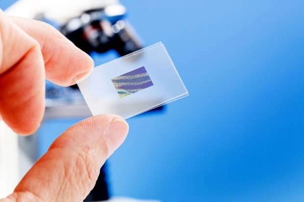
Assessing a biopsied slice of tissue to determine the grade and severity of a tumor can be highly subjective, but Stanford researchers found that computers could be trained to make accurate assessments of lung cancer tissue.
Science Photo/Shutterstock
Computers can be trained to be more accurate than pathologists in assessing slides of lung cancer tissues, according to a new study by researchers at the Stanford University School of Medicine.
The researchers found that a machine-learning approach to identifying critical disease-related features accurately differentiated between two types of lung cancers and predicted patient survival times better than the standard approach of pathologists classifying tumors by grade and stage.
“Pathology as it is practiced now is very subjective,” said Michael Snyder, PhD, professor and chair of genetics. “Two highly skilled pathologists assessing the same slide will agree only about 60 percent of the time. This approach replaces this subjectivity with sophisticated, quantitative measurements that we feel are likely to improve patient outcomes.”
The research was published Aug. 16 in Nature Communications. Snyder, who directs the Stanford Center for Genomics and Personalized Medicine, shares senior authorship of the study with Daniel Rubin, MD, associate professor of radiology and of medicine. Graduate student Kun-Hsing Yu, MD, is the lead author of the study.
Although the current study focused on lung cancer, the researchers believe that a similar approach could be used for many other types of cancer.
“Ultimately this technique will give us insight into the molecular mechanisms of cancer by connecting important pathological features with outcome data,” said Snyder.
Assessing severity of cancer
For decades, pathologists have assessed the severity, or “grade,” of cancer by using a light microscope to examine thin cross-sections of tumor tissue mounted on glass slides. The more abnormal the tumor tissue appeared — in terms of cell size and shape, among other indicators — the higher the grade. A stage is also assigned based on whether and where the cancer has spread throughout the body.
Michael Snyder
Often a cancer’s grade and stage can be used to predict how the patient will fare. They also can help clinicians decide how, and how aggressively, to treat the disease. This classification system doesn’t always work well for lung cancer, however. In particular, the lung cancer subtypes of adenocarcinoma and squamous cell carcinoma can be difficult to tell apart when examining tissue culture slides. Furthermore, the stage and grade of a patient’s cancer doesn’t always correlate with their prognosis, which can vary widely. Fifty percent of stage-1 adenocarcinoma patients, for example, die within five years of their diagnosis, while about 15 percent survive more than 10 years.
The researchers used 2,186 images from a national database called The Cancer Genome Atlas obtained from patients with either adenocarcinoma or squamous cell carcinoma. The database also contained information about the grade and stage assigned to each cancer and how long each patient lived after diagnosis.
The researchers then used the images to “train” a computer software program to identify many more cancer-specific characteristics than can be detected by the human eye — nearly 10,000 individual traits, versus the several hundred usually assessed by pathologists. These characteristics included not just cell size and shape, but also the shape and texture of the cells’ nuclei and the spatial relations among neighboring tumor cells.
“We began the study without any preconceived ideas, and we let the software determine which characteristics are important,” said Snyder, who is the Stanford W. Ascherman, MD, FACS, Professor in Genetics. “In hindsight, everything makes sense. And the computers can assess even tiny differences across thousands of samples many times more accurately and rapidly than a human.”
Bringing pathology into the 21st century
The researchers homed in on a subset of cellular characteristics identified by the software that could best be used to differentiate tumor cells from the surrounding noncancerous tissue, identify the cancer subtype, and predict how long each patient would survive after diagnosis. They then validated the ability of the software to accurately distinguish short-term survivors from those who lived significantly longer on another dataset of 294 lung cancer patients from the Stanford Tissue Microarray Database.
This brings cancer pathology into the 21st century and has the potential to be an awesome thing for patients and their clinicians.
Identifying previously unknown physical characteristics that can predict cancer severity and survival times is also likely to lead to greater understanding of the molecular processes of cancer initiation and progression. In particular, Snyder anticipates that the machine-learning system described in this study will be able to complement the emerging fields of cancer genomics, transcriptomics and proteomics. Cancer researchers in these fields study the DNA mutations and the gene and protein expression patterns that lead to disease.
“We launched this study because we wanted to begin marrying imaging to our ‘omics’ studies to better understand cancer processes at a molecular level,” Snyder said. “This brings cancer pathology into the 21st century and has the potential to be an awesome thing for patients and their clinicians.”
The work is an example of Stanford Medicine’s focus on precision health, the goal of which is to anticipate and prevent disease in the healthy and precisely diagnose and treat disease in the ill.
Stanford co-authors of the study are former postdoctoral scholar Ce Zhang, PhD; professor of pathology Gerald Berry, MD; professor of bioengineering, of genetics and of medicine Russ Altman, MD, PhD; and assistant professor of computer science Christopher Re, PhD.
The study was supported by the National Cancer Institute and the National Institutes of Health (grants U01CA142555 and 5U24CA160036-05).
Stanford’s Department of Genetics also supported the work.
-
 Krista CongerKrista Conger is a senior science writer in the Office of Communications. Email her at kristac@stanford.edu.
About Stanford Medicine
Stanford Medicine is an integrated academic health system comprising the Stanford School of Medicine and adult and pediatric health care delivery systems. Together, they harness the full potential of biomedicine through collaborative research, education and clinical care for patients. For more information, please visit med.stanford.edu.

