October 30, 2009 - By Bruce Goldman
A 2,500-year-old priest named Irethorrou will be teaching anatomy to all comers in an exhibition beginning Oct. 31. The mummified remains of this onetime inhabitant of a Middle Egyptian city will be on display in his coffin at the Legion of Honor in San Francisco, along with a reconstruct. ion of Irethorrou’s head.
The reconstruction is based on determinations of his bone structure that were part of an intensive series of state-of-the-art scans conducted in August by Stanford University School of Medicine radiologists, processed into a visual format by Stanford information technologists, and interpreted by an Egyptologist with a penchant for mummies from the town of Akhmim, the spot in ancient central Egypt where Irethorrou was found.
A Palo Alto-based software company, Fovia Inc., further wove the radiological data into a three-dimensional “fly-through” movie. Shown on a wall-mounted high-definition monitor in the exhibit gallery, the film will present visitors with visual navigations through the mummy’s anatomy, zooming in to inspect what remains of his internal organ systems and then swooping back out through the wrappings. It’s even possible to see objects, such as small amulets, buried with the mummy and hidden from view since its burial. The exhibition, “Very postmortem: Mummies and medicine,” will continue through summer 2010 at the Legion of Honor, which is part of the Fine Arts Museums of San Francisco.

The mummy that underwent a series of scans in August at Stanford is featured in an exhibition at the Legion of Honor that will run through the summer. Courtesy: Fine Arts Museums of San Francisco
Irethorrou, who has also been referred to as Iret-net Hor-iw, is one of four mummies that belong to FAMSF’s permanent collection, according to Renee Dreyfus, PhD, its curator of ancient art and interpretation. It turns out that this mummy is one of many from the same cemetery in the ancient city of Akhmim. That cemetery was excavated toward the end of the 19th century during a sudden surge in tourism to Egypt, said Jonathan Elias, PhD, director of the Akhmim Mummy Studies Consortium in Harrisburg, Pa.
Elias, an anthropologist and Egyptologist, explained that in those days, the Egyptian government was short on cash and actively sold mummies to well-heeled travelers who then brought them home, often to the United States. “There are close to 50 of them in the United States alone,” Elias said. He has tracked down a number of mummies from the Akhmim site, one of Egypt’s oldest, to analyze them for clues about ancient Egypt’s culture, diet, common diseases, average life span and the like.
“This is much easier to do if you look at many mummies from the same time and place,” said Elias, who has now participated in 20 scans of Akhmim mummies. “All the patterns just pop out at you.”
For example, Elias noted, in mummies of this era amulets are often found at the same locations on or in the body. He suspects that Egyptian physician-priests, having failed to heal a patient during life, ordered these “amulet prescriptions,” as Elias refers to them, placed in key positions as a form of magical medication to ensure that the deceased would be in top shape for the eternal afterlife.
The Stanford scans, viewed through Fovia’s software, would eventually reveal at least 14 such amulets positioned on this mummy’s body, which Dreyfus described as being “in wonderful condition.” She added, “Its state of preservation is such that all its surface bandages are still intact.”
To get the best possible results, the museum and the consortium needed a cutting-edge research scanning capacity, so they spoke to Rebecca Fahrig, PhD, associate professor of radiology. Fahrig had done this sort of thing before — namely in 2005, when a mummy from the Rosicrucian Egyptian Museum in San Jose, Calif., was brought to Stanford to be similarly scanned. But the state of the technology has advanced considerably since then, Fahrig said.
The scanning procedure, which took place on Aug. 20, required no disturbance of the mummy’s wrappings.
Using two different methods — a high-resolution CT scanner already in clinical use, and a more powerful research scanner that achieves even better resolution — Fahrig obtained some 100 billion “voxels,” the three-dimensional cubic equivalents of pixels, each measuring 0.2 micron on a side. “We went for as much CT data as we could get,” she said. “We knew we were only going to get one shot at it.”
The resulting copious data set can be accessed on demand to produce exquisitely detailed visualizations of, say, a hand or foot or head should somebody ask for it. This has applications beyond Egyptology for use in anatomical training: It’s difficult to obtain this kind of high-resolution image from a live person’s body, because the radiation would damage living tissue.

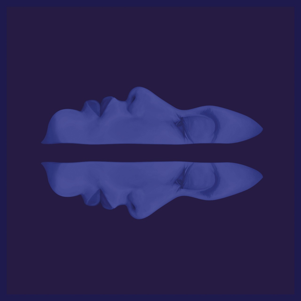
One of the scans shows the mummy's feet. Courtesy of the Fine Arts Museums of San Francisco
Chris Beaulieu, MD, PhD, chief of musculoskeletal imaging in the Department of Radiology, was one of the Stanford experts called in to check out the findings. “Many of the organs had been removed,” as is typical of mummies from that era, he said. “But it was remarkable how extremely well-preserved the mummy is in terms of its musculoskeletal structure. I could see incredible detail in the bones and joints.”
Teeth are of special interest to Paul Brown, DDS, consulting associate professor of anatomy. A dentist by training, Brown is also a researcher at the Stanford-NASA Biocomputation Center, which performed the lion’s share of the data crunching necessary to stitch the scan results into visually recognizable images.
Irethorrou’s teeth, other than being a bit worn down from the then-routine Middle Egyptian diet of stone-ground (and probably high-sand) wheat, were in good shape. He was missing just one molar.
Brown is building digital libraries of high-resolution data on teeth, hands, heads and feet for educational uses: for example, in Stanford medical training courses. Geography presents no barrier to the transmission of these three-dimensional representations, so “virtual dissections” could be performed by anatomy students anywhere in the world, said Brown.
Based on his teeth and bones, the Stanford scientists conclude that Irethorrou was perhaps 35 to 45 years old. His skeleton showed only a few traces of arthritis. He doesn’t seem to have lived a harsh life of hard labor.

The yellow spots in this scan are believed to be two of 14 amulets on the mummy that were perhaps intended to protect him in the afterlife.
The cause of Irethorrou’s death remains unknown. But the scan showed bumps on his back that were later determined, on extensive data analysis, to be lesions beneath the skin — as opposed to being simply, say, loose deposits of salt used as a desiccating agent in the embalming process — leading to speculation that an infectious disease such as a pox could have been the culprit.
Using the scan data, Elias and a colleague from DQ Models Development Group in Las Vegas reconstructed Irethorrou’s skull via rapid prototyping — a kind of 3-D printing operation whereby a shape is built up by successive layers of material deposited consecutively one above the next. Elias has since fleshed it out with clay, making sure that its thickness corresponds to meticulous measurements of the facial features of the current residents of Akhmim.
The head reconstruction will be displayed side-by-side with a bust of Ankh-Wennefer, also an Akhmim-dwelling priest who, it is believed, is Irethorrou’s father. (Writings on mummies’ coffins from this era typically state the names of the deceased’s parents.) Ankh-Wennefer’s mummy — that’s right, the mummy’s daddy’s mummy — whom Elias’s consortium had scanned in August 2008, resides in a museum in Tacoma, Wash.
“You can see the family resemblance,” said Dreyfus.
-
 Bruce GoldmanBruce Goldman is a science writer for the medical school’s Office of Communication & Public Affairs.
About Stanford Medicine
Stanford Medicine is an integrated academic health system comprising the Stanford School of Medicine and adult and pediatric health care delivery systems. Together, they harness the full potential of biomedicine through collaborative research, education and clinical care for patients. For more information, please visit med.stanford.edu.