Stanford Neurosurgical Simulation and Virtual Reality Center
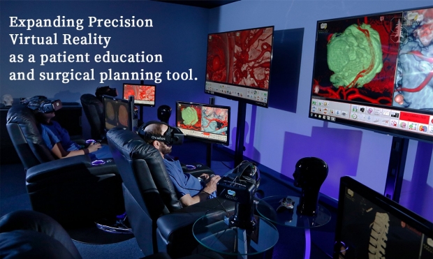

Stanford opened its Neurosurgical Simulation and Virtual Reality Center in 2016, the first institution in the greater Pacific Northwest to use patient-specific, 3-D virtual reality (VR) technology across the neurosurgery clinics, operating room, and classroom. We are also the first functional neurosurgery and spine clinics in the world to use patient-specific, 360-degree virtual reality for direct patient engagement. Since its opening, over 1,100 Stanford neurosurgery patients have had Surgical Theater 360 VR at some point during their care - from clinic consultation to preoperative planning to intraoperative navigation.
How It Works:
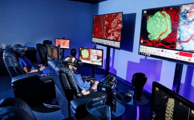
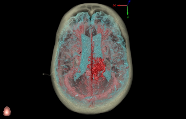
To create the 3-D images of a patient’s anatomy, scientists fuse two-dimensional images from MRI and CT scans using advanced computer programs. With the VR headsets, surgeons can then “fly” through the brain on screen – getting a close-up look at the brain tissue and vessels, all without opening up the skull.
How We Use It:
- Before and During Surgery
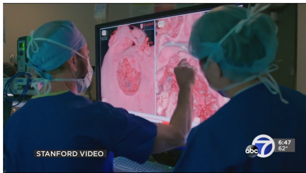
By immersing themselves in three-dimensional views of their patients’ anatomy, our doctors can more effectively plan surgical procedures. The surgeons practice the procedure using images of the actual patient, rather than generic anatomy, allowing them to map out the exact path they will take during the surgery, ahead of time.
The VR system helps surgeons in the operating room, guiding them in a three-dimensional space. The surgeons can correlate the three-dimensional images with the real-time microscopic surgical view, giving them much more detail then they would have otherwise. The three-dimensional aspect of the imagery improves accuracy during the surgery, leading to safer procedures.
- Training and Education
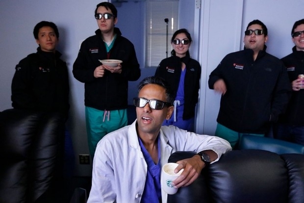
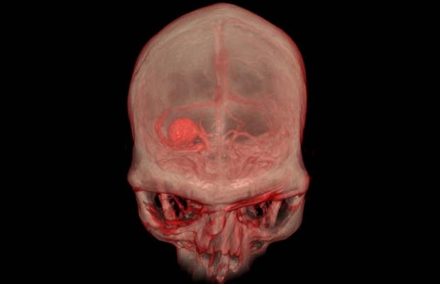
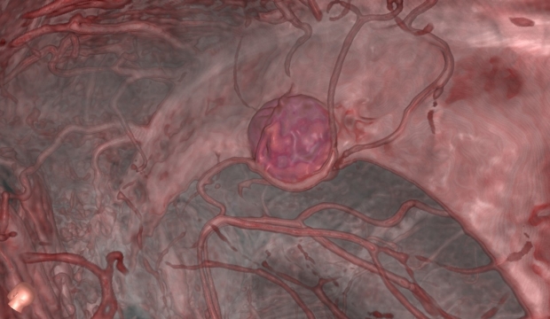
Our doctors are using this technology as an education and training tool. The system allows instructors to highlight different components of the brain, such as arteries to show an aneurysm or vascular malformation, bones to show spine and skull deformities, or tissue to show a tumor, while rotating the view to illustrate how a tumor or aneurysm appears from different angles. Students can explore complex cases together in VR and progress, as avatars, through the steps for removing a tumor or repairing an aneurysm, starting outside the skull.
- Patient Care
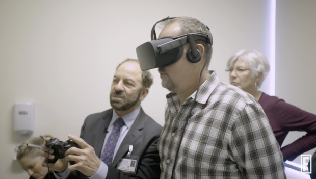
For patients, a mobile unit, complete with VR headset, can be rolled into an examination or hospital room. Being able to visualize the problem in three dimensions can help reassure patients, and is especially useful for young patients or those who don’t understand English well. Imagery can also be downloaded onto a thumb drive and given to a patient, providing a means for further review with family members.

3-D Printing
In addition to using VR technology to reconstruct 3-D images, the Stanford Department of Neurosurgery is also using 3-D printing to better understand indviduals' spine injuries, tumors, deformities, cerebrovascular disorders, aneurysms, and arterial venous malformations. Using 3-D models helps our surgeons see things they may not otherwise see when using a 2-D image on a screen, better preparing them for a surgery. Additionally, by bringing an image to life with a 3-D model, patients can view their own unique anatomy, and make more informed decisions about their treatment options.

Stanford Surgical Neuroanatomy, Fiber Tractography, and Virtual Reality Research Center
The Stanford Virtual Reality Center is a part of our Surgical Neuroanatomy, Fiber Tractography, and Virtual Reality Research Center, whose goal is to improve surgical techniques and outcomes through mastery of surgical neuroanatomy, enhancing understanding of endoscopic skull base anatomy, microsurgical neuroanatomy, and white matter dissection and imaging.
In The News
Virtual Reality Gets Real in the Operating Room
Fortune Magazine features Dr. Gary Steinberg's use of virtual reality to prepare for, and operate on a patient with an arteriovenous malformation.
New Neuroanatomy Lab Bridges Virtual Reality, Operating Room
Stanford’s Department of Neurosurgery has a new anatomy lab next door to its Neurosurgical Simulation and Virtual Reality Center. Together, the labs are a valuable resource for trainees and surgeons alike.
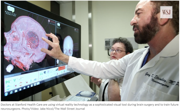
Stanford Neurosurgeons Using Virtual Reality for Training, Teaching, and Preparing for Surgery
The Wall Street Journal explores how doctors at Stanford Health Care are using virtual reality technology as a sophisticated visual tool when preparing patients, during brain surgery, and to train future neurosurgeons.
Stanford Neurosurgeons Use Virtual Reality to Show Patients Their Own Anatomy in 3D
ABC 7 Bay Area News highlights how Stanford neurosurgeons are using virtual reality to help prepare patients for brain surgery.
Virtual Reality Helps Surgeons, Reassures Patients
Stanford Neurosurgery is using a new virtual reality system to help surgeons prepare for complex surgeries and improve patients experience.
Contact Us:
Chris LeCastillo
Email: chris.lecastillo@stanford.edu
Phone: 650-725-8764


Our Stanford Neurosurgical Simulation and Virtual Reality Center has partnered with TeachAids, a pioneer in developing innovative research-based health education technologies, to create new installments of a novel concussion education program; CrashCourse.