Current Research Projects
A nanophotonic approach to building DNA using enzymatic synthesis
Punnag Padhy, Asif Mohammad, Wei Ren, Mo Wu, Bert Hesselink, Michael Jensen and Ronald Davis
Recent advances in genomics, gene editing, and synthetic biology have fueled a strong demand for higher quality, longer oligonucleotide production. However, in the past ten years, no major advances have been made in the field of solid-phase phosphoramidite chemistry, and strand length continues to be limited by its diminishing yields. Currently it costs up to $0.30 per bp to assemble a single gene ($300 / kb). This high cost is largely due to the workload of eliminating errors in assembled gene products and then choosing and sequence-validating the correct clone. Here, errors are the source of failure strands, which are invariably generated during solid-phase synthesis. Traditionally, columns and micro-wells are packaged with solid support, containing thousands of beads or glass shards, where DNA is immobilized on the surface or within channels of the support. As such, deletions are the result of 1) spent reagents and unwanted byproducts that become trapped within the support and carry over into consecutive cycles, and 2) when reagents do not completely contact all DNA molecules where beads overlap one another due to stacking. To circumvent these limitations, we propose a novel method that allows us to control the actions of an individual bead through dielectrophoresis on a plasmonic surface (Figure 1). Here, reactions are tuned to completely encapsulate each bead with minimal volume reagent droplets for high-precision synthesis. Because each bead is isolated in solution, byproducts cannot become trapped, and each has maximum contact with all synthesis reagents; it is this intimate 1:1 ratio of bead to reagent that will significantly increase the base addition efficiency allowing production of ultra-long strands of DNA > 1000 bases (). Moreover, this method will drop the cost of gene production > 100-fold (< 0.3 cents per bp) and transform research in all major fields of biomedicine.
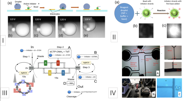
Figure 1. Schematic of nanophotonic device and synthesis approach. I) (a) simulation of bead encapsulation by reagent droplet; (b-e) microscopic view of bead encapsulation at 120V; II) (a) enzymatic synthesis conditions where TdT (terminal transferase) couples a base (with Alexa fluora) onto the initiator strands attached to the surface of the bead; (b) red filter-on, no reaction, (c) red filter-on reaction (white = fluorescence); III) enzymatic synthesis cycle on device; and IV) (A) schematic of reaction chamber on device, (B) device development at Stanford Nanofab, and (C) microscopic view of reagent droplets being injected into reaction chamber on device.
Nanoneedle diagnostic assay
A potential blood-based test for ME/CFS
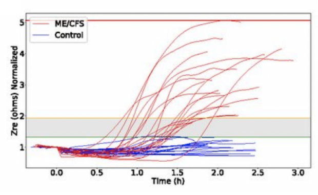
The nanoneedle biosensor is a cost-effective, versatile, ultrasensitive and high-throughput nanoelectronics device. We adapted this array to detect the electrical impedance of ME/CFS peripheral blood mononuclear cells (PBMCs). After analysis of over 20 ME/CFS patients and 20 HCs, we have established that ME/CFS PBMCs exhibit a characteristic impedance pattern when subjected to salt stress that is significantly different than controls. This work has been published in the Proceedings of the National Academy of Sciences (USA).
Further studies are underway to understand the differences between the patients’ PBMCs and those of healthy controls. To date, we know that a patient’s serum can cause HC’s PBMCs to adopt a ME/CFS impedance pattern. Preliminary fractionation experiments indicate that the factor responsible for this effect may reside in the exosome fraction of plasma. The nanoneedle assay can also be used to screen potential compounds that can revert ME/CFS impedance patterns to that of HCs.
Future efforts are being directed toward understanding the biological causes of the ME/CFS impedance pattern in the nanoneedle assay. One direction of research is to identify the specific cell population in PBMCs that may be generating the impedance pattern. Another approach is to identify the specific component(s) in patient sera that can cause HC cells to produce ME/CFS impedance patterns. In order to understand the specificity of the nanoneedle assay for ME/CFS, PBMCs from MS patients, a disease that is somewhat similar to ME/CFS, will be tested. This work has been sponsored by the Open Medical Foundation and the NIH.
Overview
Developed by Rahim Esfandyarpour, PhD, in collaboration with Ronald W. Davis, PhD, at Stanford University.
The nanoneedle biosensor is a cost-effective, versatile, ultrasensitive and high-throughput nanoelectronics device. This device measures the electrical impedance of cells from a single drop of blood.
This test is able to distinguish ME/CFS patients from healthy controls.
An analysis of over 40 ME/CFS patients and controls has established that cells from the blood of ME/CFS patients exhibit a characteristic impedance pattern when subjected to salt stress that is significantly different than the controls.
Future tests of this device will be made to:
· Determine if differences are observed between ME/CFS patients and other related diseases.
· Whether impedance signals are indicative of illness severity by testing a series of patients at altered severity.
· To test candidate drugs to see if the salt stress impedance signal can be returned to healthy control levels, revealing the potential of the nanosensor array platform for use in drug screening.
The technology will be optimized for easy clinical adoption and scaled up so that numerous FDA-approved drugs can be simultaneously screened as potential treatments
More Developing blood-based diagnostic and drug screening technology
There is currently no biological test to diagnose ME/CFS and as a result, diagnosing patients is a lengthy and costly process, constituting a fundamental impediment in patient care. This lag in diagnosis also erects barriers to research, complicating patient recruitment and potentially engaging a heterogeneous sample of patients with only superficially similar conditions.
Dr. Davis’s team is dedicated to developing inexpensive tests that can be easily used in a doctor’s office. Patients will be measured on multiple diagnostic platforms, enabling comparisons of efficacy to determine what combination of platforms would be most useful for diagnostic testing.
The Collaborative Center will continue this work to engineer a blood-based diagnostic device that would also be useful for in vitro drug screening. Dr. Davis’ team has already tested chemicals in two platforms, some of which have made the patient samples behave more like healthy samples. To validate these findings and test large numbers of samples and candidate drugs, they will further develop and optimize the technology. Eventually, the developed technology will be shared across the ME/CFS research community to accelerate its evaluation and adoption as a clinical diagnostic assay. The Stanford Genome Technology Center has developed a number of very successful diagnostic assays for other purposes that have been commercially exported and are now in clinical use. Dr. Davis’ team has experience in the complex process of developing and implementing assays that have been approved for clinical use.
Four technologies are being developed that could provide a diagnostic tool for ME/CFS:
Nanoneedle assay
Red Blood cell deformability assay
Mitochondrial function test
Magnetic levitation platform
Red blood cell deformability assay
Developing a blood-based diagnostic
In February 2019, we published in Clinical Hemorheology and Microcirculation our results that demonstrated a significant decrease in deformability of red blood cells (RBCs) from ME/CFS patients compared to HCs, which may have origins in oxidative stress. We hypothesize that altered microvascular perfusion due at least in part to reduced RBC deformability can be a possible cause for ME/CFS symptoms. Our data also suggest that RBC deformability may serve as a potential biomarker for ME/CFS, albeit further studies are necessary for non-specific classification of the disease.
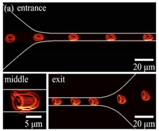
We have assembled a collaborative team to work on several approaches to studying RBC deformability in ME/CFS patients. In addition to Ron Davis and personnel at the Collaborative Research Center at Stanford, three other PIs and their labs are involved: Anand Ramasubramanian of San Jose State, who was one of the corresponding authors of the RBC deformability publication; Juan Santiago, director of the Stanford Microfluidics Laboratory; and Eric Shaqfeh, a Stanford expert in simulation and analysis of blood flow in microfluidics. Together the group has designed and fabricated 10 design variations for a microfluidic platform that can yield imaging data suitable for comparison to computer simulations generated by the use of High-Performance Computing Resources by the Shaqfeh group. Microfluidic devices suggested from the simulations will be tested with the ultimate goal being the creation of a sensitive diagnostic device. This project is sponsored by the Open Medical Foundation.
Overview
Developed by Mohsen Nemat-Gorgani, PhD, of Stanford University, and Anand Ramasubramanian, PhD, of San Jose State University. In collaboration with Juan Santiago, PhD, and Eric Shaqfeh, PhD, and Ronald W. Davis, PhD, at Stanford University.
Several studies have implicated a role of oxidative stress in ME/CFS. Red blood cells (RBCs) are potent scavengers of oxidative stress and their shape changes appreciably in response to oxidative stress; this has been observed in certain inflammatory conditions including obesity and diabetes.
The shape of RBCs determine how well these cells can move through blood vessels so it seems pertinent to determine if RBCs in ME/CFS patients are affected. This has led to the development of a microfluidic device that mimics blood flow through microcapillaries.
Preliminary studies have shown that RBCs from ME/CFS patients had an altered rate of movement through microcapillaries and that RBCs from ME/CFS patients had reduced deformability.
The significant decrease in deformability of RBCs from ME/CFS patients may have origins in oxidative stress and suggests that altered microvascular perfusion can be a possible cause for ME/CFS symptoms.
This work has been accepted for publication in Clinical Hemorheology and Microcirculation and also has been accepted as an abstract for the American Society of Hematology 60th Annual Meeting.
Mitochondrial function test
Developed by Julie Wilhelmy, PhD, in collaboration with Ronald W. Davis, PhD, at Stanford University.
The Seahorse instrument measures oxygen during energy production processes that occur in the mitochondria. A protocol using the Seahorse has revealed a significant difference between activated T-cells of ME/CFS patients and healthy controls.
The instrument is commercially available, which will allow other laboratories to easily reproduce the results.
Next-generation tools for metabolic and protein engineering in S. cerevisiae
Multiplexed accurate genome editing with short, trackable, integrated cellular barcodes (MAGESTIC).
We have developed several next-generation tools that leverage advances in synthetic biology and CRISPR/Cas9 genome editing. These tools include a core technology called Recombinase Directed Indexing (REDI), which transforms complex oligonucleotide libraries into high-density ordered arrays of yeast clones, each containing a unique sequence-verified oligonucleotide of interest. In addition, Multiplexed Accurate Genome Editing with Short, Trackable, Integrated Cellular Barcodes (MAGESTIC) enables high-throughput modification of the yeast genome at unprecedented scale and precision. We are applying these tools to produce new natural product chemicals by expressing biosynthetic pathways from other organisms in an optimized yeast host. For example, heterologous expression of several plant genes, combined with modifications to native yeast metabolism, is being used to produce the natural product Strictosidine, a common precursor to several medically and commercially valuable chemical entities that is extremely expensive to isolate from natural sources. We are also applying our technologies towards engineering of proteins. Projects include systematically evaluating the effects of synonymous genetic changes on translation efficiency, mapping amino acids substitutions that alter drug binding and resistance, and engineering novel enzymatic activities of commercial value. This work is sponsored by the NIH.
T cells and the molecular immunology of ME/CFS
T cell diversity in ME/CFS
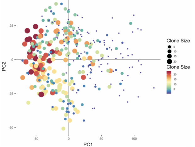
Our immune system consists of a vast repertoire of T cells, with an estimated diversity of over 108 T cell receptor (TCR) beta chains alone, permitting the detection of many different foreign antigens. Upon recognition of an infected cell, a T cell expands clonally, retaining its original TCR sequences, which are so unique they act as a barcode. While TCR repertoires have been investigated in ME/CFS using bulk RNA-Seq, differences in activated T cells (which constitute only 2–5% of this population) would be easily obscured by this approach. We have developed a robust single-cell method for TCR sequencing and phenotyping. This effort is sponsored by the Open Medical Foundation and the NIH.
Overview
This study is led by Michael Sikora, in collaboration with Mark M. Davis, PhD, Lars M. Steinmetz, PhD, and Ronald W. Davis, PhD, at Stanford University.
Beginning in 2016, the aim of this study was to establish the role of T cells and the immune system in ME/CFS by examining the genetic material in T cells - immune cells that identify and kill infected cells. The rationale is that aspects of of genetic material in these cells can inform whether they are actively fighting an infection and potentially what the cause of the infection is.
Many studies have shown that the immune system is affected in ME/CFS patients, e.g., low activity of NK cells, altered levels of cytokines (signaling molecules of the immune system), and the likelihood of a microbial infection preceding the illness. None of these studies are yet to implicate T cells or define their activity in ME/CFS patients.
The investigation of the immunological basis of ME/CFS will have several parts that overall will help determine if ME/CFS is an autoimmune disease and what immune factors may be triggering ME/CFS or sustaining it as a chronic disease.
Dr. Mark Davis’ team is investigating the clonal expansion of T cells in ME/CFS, including what they might be targeting – viruses, bacteria, or self (autoimmune).
Dr. Ron Davis’ team has invented a highly accurate, cost-effective method for HLA gene sequencing and a very sensitive method for detecting viral DNA as a sign of viral infections, which he is using in this project.
Dr. Lars Steinmetz’ team has developed effective methods for sequencing RNA from single T cells, which they are using to understand how T cell behavior may be different in ME/CFS.
Metabolic Trap Hypothesis
IDO
The hypothesis that environmental stressors interact with a potentially common genetic lesion led to the discovery of common, damaging mutations in the IDO2 gene in the Severely Ill Patient (SIP) cohort. These mutations unmask the well-known substrate inhibition of its partner enzyme, IDO1. This nonlinear dependence of IDO1 on its amino acid substrate, tryptophan (Trp), opens the door to what, in the language of nonlinear systems theory, is called a bistable system, which can be forced to shift by environmental stimuli to a stable pathological steady state. This work is funded by the Open Medical Foundation.
Overview
This study is led by Robert D. Phair, PhD, of Integrated Bioinformatics, Inc, in collaboration with Julie Wilhelmy, PhD, and Ronald W. Davis, PhD, at Stanford University.
Beginning in 2018, the aim of this study was to test the hypothesis developed by Dr Phair that a crucial component of metabolism in ME/CFS patients appears to be “trapped” in an unhealthy state.
The metabolic trap theory emerged from the genetic and metabolomics data from the Severely ill Patients Study (SIPS). Using previously published work, Dr Phair has developed a computational program that can model the flow of metabolism throughout the body and its cells. Using this technology, Dr Phair can determine any points that may disrupt the flow using genomic and metabolic information and this has led to the hypothesis of a metabolic trap that occurs in ME/CFS patients at the point in metabolism where tryptophan is converted to serotonin and kynurenine.
To validate the hypothesis, experiments were initiated to measure the levels of tryptophan and kynurenine in cells from ME/CFS patients. This is a complicated procedure as taking cells out of the body without disrupting their inherent metabolism is fraught with problems.
Metallomics in ME/CFS patients
Chronic Fatigue Syndrome, or Myalgic Encephalomyelitis (ME/CFS) is a chronic debilitating condition that is characterized by fatigue, post-exertional malaise, unrefreshing sleep, and either cognitive impairment or orthostatic intolerance. The critical barrier to progress in this field is that Chronic Fatigue Syndrome is diagnosed based on subjective symptoms, rather than specific, quantifiable, and actionable biomarkers. The lack of objective biomarkers raises the possibility that Chronic Fatigue Syndrome is a disease with several different etiologies. Although the cause(s) of ME/CFS remain unclear, there are several non-specific indications of organic disease that differentiate patients from healthy controls, including increased levels of oxidative and nitrosative stress, neuroinflammation, immune system abnormalities, and disturbed energy metabolism. Notably, a recent study evaluating thyroid function in ME/CFS patients identified a subset of individuals with “Low Free T3 Syndrome,” which is characterized by a reduced conversion of tetraiodothyronine (thyroxine, T4) to triiodothyronine (T3).
In order to address whether environmental factors contribute to a potential disease mechanism in ME/CFS, we performed a metallomics analysis of hair from 47 patients and 31 healthy controls. We identified a subset of patients with hair mercury levels exceeding the World Health Organization (WHO) reference range of 2.0 ugHg/g hair. Heavy metals, especially methylmercury, exert toxic effects by directly inhibiting various enzymes, but these metals also have a strong affinity for the essential trace metal, selenium. It is possible that mercury exposure reduces the bioavailability of selenium, even when concentrations of selenium in tissues do not appear to be limiting. The significance of this observation is that selenium is a required cofactor for thyroid hormone metabolism and management of antioxidant defenses, which have both been shown to be deficient in patients diagnosed with ME/CFS. Our central hypothesis is that environmental exposure to methylmercury reduces the bioavailability of selenium, and causes a reduction in various selenoenzyme activities in patients with chronic, unexplained fatigue. This project is sponsoered by the Open Medical Foundation.
ME/CFS severely ill big data study
Killer cell immunoglobulin-like receptors (KIRs)
The severely ill patient (SIP) Big Data study continues to yield intriguing insights into the pathology of myalgic encephalopathy/chronic fatigue syndrome (ME/CFS). Natural killer (NK) cells are an immune cell subtype implicated in this disease, and recent analysis of the SIPs data has found polymorphisms in a complex family of receptors expressed by NK cells that are in association with ME/CFS. This family of receptors is called killer cell immunoglobulin-like receptors (KIRs), and they regulate NK cells by binding to major histocompatibility complex proteins, which are encoded by the human leucocyte antigen (HLA) system and are central to human immunity. To date, five patient-enriched variants in the KIR genes (3DL3, 2DL4 and 3DL2) have been found in association with ME/CFS. However, both the HLA and KIR genes are challenging to sequence due to sequence diversity as well as homologous and repetitive regions. To meet this challenge, Chia-Jung Chang and Wenzhong Xiao have collaborated with Marcelo Fernandez Vina, an expert in clinical HLA sequencing at Stanford, to establish new standards for HLA and KIR sequencing so that the potential role of NK cells can be better understood in ME/CFS by sampling a larger number of ME/CFS patients. This research is funded by the Open Medical Foundation.
Overview
This study is co-led by Ronald W. Davis, PhD, and Wenzhong Xiao, PhD.
The aim of the Severely Ill Patient Study (SIPS), which started in 2015, was to conduct a comprehensive “Big Data” analysis on severely ill ME/CFS patients in order to begin an exploration to find the molecular basis of ME/CFS. The rationale of the study was that molecular differences would show the strongest signal in severe cases of the illness when compared to healthy controls.
Over 1,000 tests have been conducted per patient, which is the largest (to our knowledge) dataset ever generated in any patient.
Using samples of saliva, blood, urine and feces, this big data study examined the following:
Genome
Gene expression on individual immune cell types
Metabolomics
Microbiome
Cell-free DNA sequencing and quantitation
Cytokines
Clinical tests
The value of a study of this magnitude is that it provides a foundation of invaluable information to generate hypotheses and to develop diagnostics and treatments. Further, all samples from one patient are taken at one point in time, enabling all measurements to reflect exactly what was happening in the body at that one time point. This allows the possibility for the discovery of biomarkers involving the relationships between different body systems that would not ordinarily be found.
This large dataset has been made available online to all interested researchers in the ME/CFS Data Center at Stanford, with the purpose of inspiring research globally, eliminating duplication of effort, and building a collaborative network of scientists.
Extended big data study
Killer cell immunoglobulin-like receptors (KIRs)
This study is led by Fereshteh K. Jahaniani, PhD, in collaboration with Michael P. Snyder, PhD, and Ronald W. Davis, PhD, at Stanford University.
Beginning in 2016, the aim of this study was to extend the Severely ill Patient Study (SIPS) and conduct a comprehensive “Big Data” analysis on ME/CFS patients and their families. The rationale of this study is that by using the unaffected family members as a control, the differences observed in the ME/CFS patients will be more specific to the disease and less related to differences in genetics, environment, and diet.
Almost as extensive as the SIPS study, blood from each patient is being used to examine the following:
· Genome
· Gene expression
· Metabolomics
· Proteomics
· Cytokines
By comparing patients to healthy blood relatives, we are more likely to understand what genes cause or contribute to the development of ME/CFS. This data will also be integrated with the SIPS data.
The team anticipates that this study will help define meaningful subgroups of patients, biomarkers that could be useful in diagnosis and monitoring of disease progression, and molecular defects that could be targeted with new treatments.