CME Radiology Grand Rounds
When:
2nd & 4th Fridays, 12:00pm - 1:00pm
Exceptions:
5/3/24 & 5/17/24 will be the 1st & 3rd Friday of the month.
The Moskowitz Lectureship will be on the first Thursday, 11/2/23, from 5:30-6:30pm.
Where:
Li Ka Shing Learning and Knowledge Center & Zoom.
Zoom link:
https://stanford.zoom.us/j/91767596068?pwd=aTRUVEc3K28wcHFsQ3UxUXQ5T05Hdz09
1. Critically analyze research, guidelines and appropriate use criteria to develop best-practice diagnosis and treatment strategies
2. Evaluate latest innovations in imaging to assess safety and effectiveness
Course Director: H. Henry Guo, MD, PhD
Course Admin: Susan Hanlock
Accreditation
The Stanford University School of Medicine is accredited by the Accreditation Council for Continuing Medical Education (ACCME) to provide continuing medical education for physicians.
Credit Designation
The Stanford University School of Medicine designates this live activity for a maximum of: 1.00 AMA PRA Category 1 Credit(s)™. Physicians should claim only the credit commensurate with the extent of their participation in the activity.
Cultural and Linguistic Competency
California Assembly Bill 1195 requires continuing medical education activities with patient care components to include curriculum in the subjects of cultural and linguistic competency. The planners and speakers of this CME activity have been encouraged to address cultural issues relevant to their topic area. The Stanford University School of Medicine Multicultural Health Portal also contains many useful cultural and linguistic competency tools including culture guides, language access information and pertinent state and federal laws. You are encouraged to visit the portal: http://lane.stanford.edu/portals/cultural.html.
CME Radiology Grand Rounds 2022 - 2023
CME Radiology Grand Rounds 2021 - 2022
CME Radiology Grand Rounds 2020 - 2021
CME Radiology Grand Rounds 2018 - 2019
CME Radiology Grand Rounds 2017 - 2018
CME Radiology Grand Rounds 2016 - 2017
CME Radiology Grand Rounds 2015 - 2016
CME Radiology Grand Rounds 2014 - 2015
CME Radiology Grand Rounds 2013 - 2014
CME Radiology Grand Rounds 2012 - 2013
CME Radiology Grand Rounds 2011 - 2012
2023 - 2024 Schedule
September
Friday, September 22, 2023
12:00-1:00PM | LKSC 120 & Zoom
Sanjiv Sam Gambhir Lectureship

Carolyn Bertozzi, PhD
Baker Family Director of Stanford Chem-H, Ann T. and Robert M. Bass Professor in the School of Humanities and Sciences and Professor, by courtesy, of Chemical and Systems Biology and of Radiology
Stanford University
Bioorthogonal Chemistry, from Basic Science to Clinical Translation
Bioorthogonal chemistry has opened new avenues for biological research and therapeutic science. Here I will present the origin story of bioorthogonal chemistry as a tool for molecular imaging of cell surface glycans, advances in reaction development, and recent translational work leading to clinical drug candidates.
Learning objectives:
- Understand the concept of bioorthogonal chemistry
- Learn about applications in the biopharma industry
October
Friday, October 13, 2023
12:00-1:00PM

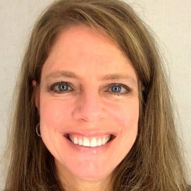
Katherine Maturen, MD, MS
Clinical Professor, Radiology
Associate Chair, Ambulatory Radiology and Strategy
Medical Director, East and Northeast Radiology Ambulatory Care Units
Michigan Medicine, University of Michigan
Ovarian Cancer for Radiologists: How We Can Do Better
We will discuss fundamentals of ovarian cancer pathophysiology, demographics and risk, as well as imaging characterization of adnexal masses and staging of ovarian cancer. Research gaps and opportunities for improvement in imaging care will be highlighted throughout the presentation.
Learning Objectives: At the completion of this session, participants will: Understand the tubal origin of most ovarian cancers and ramifications for imaging Recognize high risk features in adnexal masses on US and MRI Identify disease sites that limit resectability in staging CT for ovarian cancer Commit to being relentless detectives for their patients with suspected cancer or recurrence
Friday, October 27, 2023
12:00-1:00PM
Donna Roberts, MD, MS
Professor
Department of Radiology and Radiological Sciences
Department of Neurosciences, Neuroscience Research
Research Director, Division of Aerospace Medicine
MUSC Campus Director, SC Space Grant Consortium
Medical University of South Carolina
The Effects of Spaceflight on Astronaut Brain Health as Indicated on MRI with Translation to Terrestrial Clinical Populations
Objectives:
- Describe the effect of spaceflight on human brain structure as determined by MRI.
- Describe the relationship between structural brain changes and changes in cognitive and motor performance in astronauts during long-duration spaceflight.
- Describe how studying the adaptation of the brain to spaceflight and hydrocephalus associated with long-term spaceflight (HALS) may contribute to the understanding of disorders of cerebral spinal fluid (CSF) homeostasis on Earth.
CSF homeostasis is complex and incompletely understood. My group and others, have documented changes in brain structure in astronauts following spaceflight including upward shift of the brain, ventricular enlargement, and enlargement of the perivascular spaces. These structural changes are greater than would be expected for normal aging and occur in astronauts following long-term missions (>3 months) on the International Space Station, but not in astronauts who flew short-duration missions (1-2 weeks) on the Space Shuttle. Remodeling of cerebral structures in response to abolishment of the habitual gravitational stress of Earth implies a previously unrecognized gravitational dependency. Further investigation of the impact of gravity on CSF dynamics may hold significance not only for astronauts but may also be applicable to understanding the underlying etiology of CSF disorders on Earth.
November
Thursday, November 2, 2023
5:30-6:30PM | LK120 & Zoom
Etta K. Moskowitz Lectureship

Deborah Liu, MBA
Chief Executive Officer of Ancestry.com
Fireside Chat
Abstract coming soon.
Friday, November 10, 2023
12:00-1:00PM | LK101 & Zoom
Diversity Lecturship

Vaz Zavaletta, MD, PhD
Assistant Professor of Radiology (Pediatric Interventional Radiology)
Children's Hospital of Colorado
Seeing the Full Spectrum: Gender Inclusive Care in Radiology
LGBTQIA+ people face lower healthcare utilization rates due to discrimination, poor experiences in healthcare, and barriers to accessing care. There is an increasing need to improve care and reduce health care disparities for the LGBTQIA+ population. The medical community can begin by educating themselves on LGBTQIA+ terminology, using inclusive language and developing cultural competence in clinical settings. In order to achieve this it is first important to understand that Sex and Gender are distinct and that both Sex and Gender exist on continuums. By thoughtfully incorporating this knowledge into our research and clinical practice, the radiology community will enhance the healthcare experiences of all patients.
Learning Objectives:
- Define concepts and terminology regarding gender diversity within a gender expansive (non-binary) framework.
- Recognize the unique health care experiences of a gender diverse individuals and identify barriers to equitable health care.
- Propose strategies and solutions to create a more inclusive and welcoming environment for gender diverse individuals in healthcare.
December
CANCELLED - Friday, December 8, 2023
12:00-1:00PM | LK120 & Zoom
Cancelled
January
Friday, January 12, 2024
12:00-1:00PM | LK101 & Zoom
Juergen K. Willmann Lectureship
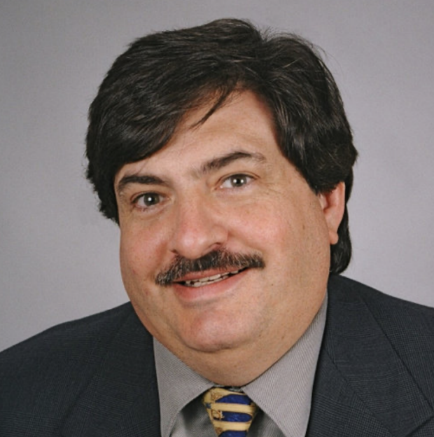
Richard Barr, MD, PhD
Professor of Radiology, Northeastern Ohio Medical University
Southwoods Imaging
Editor-in-Chief Journal of Ultrasound in Medicine
Multiparametric Ultrasound for Chronic Liver Disease
Chronic liver disease is a worldwide epidemic with estimates of 25-30% people affected. MAFLD (NAFLD) is now the major cause of chronic liver disease. With the large number of affected individuals an inexpensive, safe, rapid, accurate method of evaluation is need to not only make the diagnosis but also to follow-up with patients on treatment. Ultrasound is uniquely able to fulfill these requirements. This lecture will review the use of multiparametric ultrasound including liver stiffness estimation, liver fat estimation, and new techniques that may provide some information on inflammation.
Learning Objectives:
- Review the use of ARFI based techniques for estimation of liver stiffness
- Discuss the use of quantitative ultrasound techniques for liver fat estimation
- Present how to use Multiparametric Ultrasound in Clinical Practice
Friday, January 26, 2024
12:00-1:00PM | LK120 & Zoom
Etta K. Moskowitz Fund Resident Research Awards
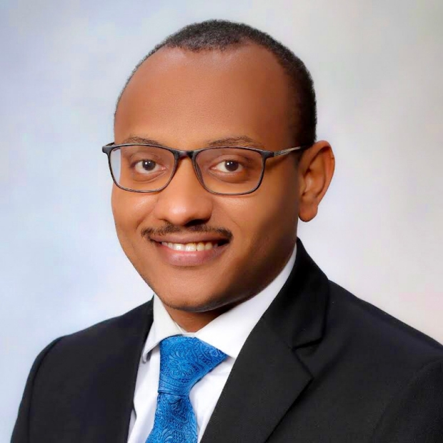
Mohamed Muneer, MD
Resident in Radiology, Stanford University
Reflector (Scout) localization at the time of breast biopsy instead of a biopsy clip: an academic center experience
Preoperative image-guided localization of non-palpable breast findings is pivotal in reducing the need for reoperation by ensuring a negative resection margin and yielding improved cosmetic and patient satisfaction outcomes. Wire-guided localization (WGL) has historically been the gold standard, but emerging alternatives, such as wireless-guided localization (Wi-GL), offer distinct advantages.
Wi-GL stands out by decoupling the localization procedure from surgery, allowing for more flexible surgical scheduling. While radioactive seed was the first commercially available Wi-GL option, it requires strict regulatory control, with careful tracking from seed deployment to retrieval. Radar localization devices, such as the reflector (Scout®) provide additional convenience without the regulatory burden. FDA approval in 2017 of the Scout for long-term use enabled the placement of the reflector at the time of biopsy, eliminating the need for a separate localization procedure for surgically intervened lesions. However, considerations arise regarding the permanent presence of the reflector if surgery does not proceed, impacting future imaging (e.g., MRI) and localization. Also, if the patient undergoes a mastectomy, then localization is essentially not needed, given the relatively higher cost of Wi-GL as compared to WGL or no localization.
Currently, there is little guidance on choosing between wireless devices and biopsy clips during breast biopsy. The study retrospectively evaluates eventual surgical utilization of reflectors placed at the time of biopsy, aiming to identify factors influencing successful utilization. The forthcoming presentation will shed light on these factors and contribute to refining the decision-making process for choosing reflector devices or biopsy clips during breast biopsy.
Mentor: Dr. Brittany Dashevsky MD, DPhil

Joy Wu, MD, MPH
Resident in Radiology, Stanford University
Eye-tracking assisted target tumor quantification on FDG-PET/CT scans.
FDG-PET/CT studies are the standard of care for staging and following up of multiple cancers. Currently, assessing disease progression involves time consuming manual measurements of multiple index tumor lesions. Many have attempted building stand-alone AI models to automate this. However, these models often fail when encountering rare long tail cases in real clinical workflow. With advances in eye tracking hardware, we attempt to harness the fastest muscles in the human body in a Human Computer Interaction system for tumor quantification. We show that our baseline algorithm is on average 84% successful in utilizing insights gathered from expert gaze attention and features from PET images for measuring target hypermetabolic lesions.
Mentors:
Dr. Guido Davidzon
Dr. Curtis Langlotz
Davis Vigneault, MD, DPhil
Resident in Radiology, Stanford University
A Novel Neural Network Architecture for Native Resolution AAA Segmentation
Abdominal aortic aneurysm (AAA) is common and asymptomatic throughout its disease course, but carries the risk of catastrophic rupture and hemorrhage. Highly accurate aortic segmentation is critical to avoid treatment errors near the treatment threshold as well as in the assessment of candidate pharmacological therapies. Previously published AAA segmentation models suffer from some combination of brittle preprocessing procedures, failure to account for global context, unacceptable downsampling and consequent loss of spatial resolution, lack of end-to-end training, or inadequate validation.
We propose a novel convolutional neural network (CNN) architecture which addresses all major limitations of the prior work. The proposed architecture involves three sequential modules responsible for coarse segmentation, transformation, and fine segmentation, respectively. 71 patients with small AAA enrolled in the LIMIT trial who have undergone baseline abdominopelvic CTA were included. Training and testing were performed using an 80:20 split.
The proposed model produces high-quality, native resolution, volumetric AAA segmentations. Ablation experiments demonstrating the effect of architectural choices and training techniques are performed.
We have presented a novel neural network architecture capable of providing high quality, native-resolution AAA segmentations. The segmentations produced will be valuable both for clinical practice and for assessment of candidate pharmacological therapies. Simple modifications to the proposed structure are likely to be valuable in other tasks where high-resolution segmentations of small structures are required.
Mentor: Dr. Dominik Fleischmann
February
Friday, February 9, 2024
12:00-1:00PM | LK120 & Zoom

Sylvia Plevritis, PhD
Professor of Biomedical Data Science and of Radiology (Integrative Biomedical Imaging Informatics at Stanford)
Stanford University
Dissecting the Tumor Microenvironment with AI/ML Tools
Friday, February 23, 2024
12:00-1:00PM | Zoom only

Elizabeth Morris, MD
Professor and Chair
Department of Radiology
UC Davis Health
New Ways of Thinking about Breast Cancer Screening
March
Friday, March 8, 2024
12:00-1:00PM | LK130 & Zoom
Fabrice Lucien-Matteoni, PhD
Assistant Professor of Immunology & Urology
Mayo Clinic
Extracellular vesicles as non-invasive biomarkers for the management of prostate cancer
Extracellular vesicles (EVs) have emerged as promising biomarkers for cancer detection due to their ability to carry molecular cargo, including DNA, RNA, and proteins, reflecting their cell of origin. This presentation highlights the clinical relevance of tumor-derived EVs in the management of prostate cancer. We will discuss the characteristics of tumor-derived EVs and their potential as diagnostic and prognostic biomarkers in prostate cancer. Furthermore, we will explore how integrating EV analysis with imaging techniques can enhance prostate cancer diagnosis and risk stratification. Additionally, we will address the technical challenges associated with EV purification and profiling. This discussion aims to underscore the significance of EVs as a non-invasive tool in the comprehensive management of prostate cancer.
Session Learning Objectives:
Upon conclusion of this activity, participants will be able to:
- To describe basic principles of extracellular vesicles as non-invasive biomarkers
- To summarize current state of extracellular vesicle-based liquid biopsies in prostate cancer
- To identify opportunities for integrating liquid biopsy with imaging in oncology
Friday, March 22, 2024
12:00-1:00PM | LK101 & Zoom
Donald Resnick, MD
Professor Emeritus
Department of Radiology
University of California, San Diego
Of Skeletons, Scholars, and Sleuths: A Look Back, A Look Around, and A Quick Look Ahead
The history of musculoskeletal imaging at UCSD will be reviewed with emphasis on the importance of imaging-pathologic correlation. Some predictions for the future will also be introduced.
Learning objective
To illustrate the importance of imaging-pathologic correlation, especially with regard to degenerative and inflammatory disorders of the vertebral column
April
Friday, April 12, 2024
12:00-1:00PM | LK120 & Zoom
Lisa Bodei, MD, PhD
Attending, Director, Targeted Radionuclide Therapy
Department of Radiology
Molecular Imaging and Therapy Service
Professor of Radiology, Weill Cornell Medical College of Cornell University
Memorial Sloan Kettering Cancer Center
Theranostics, the age of Enlightenment
Radiotheranostics are a unique type of cancer drugs which enable simultaneous imaging and therapy. Theranostics can be used to visually assess the biodistribution of the radiopharmaceutical and for the selection of patients to receive the therapeutic drug (which can be described as ‘seeing what you treat’). Thus, theranostics help reducing the risks of failure associated with drug development, by allowing the visualization and quantification of both the presence and engagement of the target. Old and recent clinical successes achieved thus far, particularly in neuroendocrine tumors and prostate cancer, go in parallel with the development of challenges associated with the effective and widespread deployment of new agents, as well as their future potential and emerging opportunities.
One of the major challenges in this field is the relatively short duration of treatment response. Several new strategies to improve the efficacy and tolerability of radiotheranostics have been proposed. Among these, the use of alpha emitters is very promising to overcome the intrinsic radioresistance of cancer cells. Also, equally important, is the individualization treatments based on the specific dosimetric estimates for the tumor and normal organs, as well as the determination of tissue radiosensitivity.
Learning objectives
On successful completion of this activity, participants should be able to describe
- The application of theranostics in neuroendocrine tumors and prostate cancer.
- The approved radiopharmaceuticals and their efficacy and tolerability profile.
- New strategies for the improvement of the treatment response.
Friday, April 26, 2024
12:00-1:00PM | LK130 & Zoom

Sanjeev Bhalla, MD
Section Chief, Cardiothoracic Imaging
Professor of Radiology, Cardiothoracic Imaging
Assistant Residency Program Director
Vice-Chair for Education
Mallinckrodt Institute of Radiology
Going for Broke: Imaging of Cardiothoracic Trauma
This seminar will focus on the imaging findings in cardiothoracic trauma with emphasis on a location based approach and the use of direct and indirect signs. Penetrating trauma will also be discussed.
Session Learning Objectives
- Highlight the concept of direct vs indirect signs in Cardiothoracic Trauma Imaging
- Discuss the understanding the value of points of differential deceleration
- Emphasize how blunt and penetrating trauma may vary in their appearances
May
CANCELLED - Friday, May 3, 2024
12:00-1:00PM | Room TBD & Zoom
Cancelled
Friday, May 31, 2024
12:00-1:00PM | LKSC120 & Zoom
Gary M. Glazer Lectureship
Geoffrey Rubin, MD, MBA
Professor and Chair
Department of Medical Imaging
College of Medicine – Tucson
University of Arizona
TBD
Abstract coming soon.