A circadian code controls the switch that produces fat cells, according to a new study by Stanford researchers.
April 3, 2018 - By Rosanne Spector
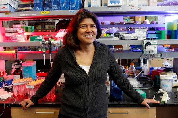
Mary Teruel and her colleagues discovered that rising and falling levels of hormones known as glucocorticoids can affect weight gain.
Paul Sakuma
New research provides the first molecular understanding of why people gain weight due to chronic stress, disrupted circadian rhythms and treatment with glucocorticoid drugs: It’s all in the timing of the dips and rises of a class of hormones called glucocorticoids — predominantly the “stress hormone” cortisol, according to a new study by Stanford University School of Medicine researchers.
The research suggests new strategies to reduce weight gain by controlling the timing of hormonal pulses, said Mary Teruel, PhD, assistant professor of chemical and systems biology and senior author of the study, which was published online April 3 in Cell Metabolism.
“It explains why treatments with glucocorticoid drugs, which are often essential for people with rheumatoid arthritis and asthma to even function, are so linked with obesity, and it suggests ways in which such treatments can be given safely without the common side effects of weight gain and bone loss,” she said.
Fat cells normally turn over at a rate of 10 percent per year; they die and are replaced by newly differentiated fat cells. What has long fascinated Teruel is how we stay at this rate, and the mystery of what flips the switch that leads to weight gain.
“Now we know the circadian code that controls the switch, and we’ve identified key molecules that are involved,” Teruel said.
The team found that fat-cell maturation ramps up if the trough in exposure to glucocorticoids lasts less than 12 hours.
Teruel and her team made these findings during their effort to identify the molecular mechanism that precursor fat cells use to sense and filter out short pulses and normal oscillations of glucocorticoids.
The 24-hour hormone cycle
A healthy person’s level of glucocorticoids rises and falls in a circadian 24-hour cycle, peaking around 8 a.m., dropping to its lowest around 3 a.m. the next day, and then rising back to its peak about five hours later. The rise is a wake-up signal that gets us moving and turns on our appetites. The glucocorticoid levels in our bloodstream are also increased by stress — short spikes are induced by short-term stress, such as exercise, and sustained levels are caused by chronic stress.
Researchers have long known that glucocorticoids trigger precursor cells to convert to fat cells, and that our fat tissue contains a huge excess of precursor cells that could convert, given the right signals. Under healthy conditions, less than 1 percent of a person’s precursor fat cells are converting into fat cells. This low rate of conversion is essential for replacing damaged mature cells and for renewing and maintaining healthy fat tissue.
Yes, the timing of your stress does matter.
Hence Teruel’s puzzlement: “So what stops normal, healthy daily increases in our glucocorticoid levels due to circadian rhythms and healthy short-term stresses from causing all our precursor cells to convert into fat cells? Why aren’t we drowning in fat every time glucocorticoid levels go high in the morning due to normal circadian rhythms or when our glucocorticoid levels spike when we exercise or go from a warm building out into the cold? And why is losing the normal rhythm of glucocorticoid secretion — such as in conditions of chronic stress, jetlag and sleep disruption in shift-workers — so linked to obesity?”
The timing of glucocorticoid pulses had not been studied before, but Teruel thought it might offer the answer.
In the first of the series of experiments, graduate students Zahra Bahrami-Nejad and Michael Zhao, co-lead authors of the study, worked around the clock to expose precursor fat cells to glucocorticoids in carefully timed pulses over the course of four days, alternately bathing the petri-dish-grown cells in fluids with and without glucocorticoids and assuring that the total exposure to the hormone remained the same. They stained and imaged the cells so they could count how many of the precursor cells matured into fat cells. They found that one pulse of glucocorticoids lasting 48 hours led most of the cells to differentiate, while shorter pulses with at least 12 hours between them resulted in minimal differentiation.
To uncover how the precursors are able to sense the duration of the hormonal pulses and filter out short pulses, the researchers used single-cell live imaging to track PPAR-gamma protein levels in thousands of individual cells over several days while the precursors became fat cells. PPAR-gamma is a protein that correlates with the fat cell’s maturity: When PPAR-gamma levels increase to a certain threshold level in a fat precursor cell, the precursor cell will convert in to a fat cell. Leading up to this experiment, Bahrami-Nejad had worked for about two years using CRISPR gene-editing technology to attach a fluorescent probe to all of the PPAR-gamma proteins the precursor fat cells produced. By measuring the fluorescence, she and Zhao were able to quantify levels of PPAR-gamma produced in the cells, which enabled them for the first time to watch individual cells convert from precursor cells into fat cells as it happened.
Relying on two types of feedback
The researchers’ experiments and computer modeling indicated that the system must rely on two types of positive feedback — fast and slow — to enable precursor cells to ignore the normal rise and fall of glucocorticoids, as well as short daytime pulses, yet respond to long pulses. Their work had previously found that a protein called CEBP-alpha provides fast positive feedback — meaning PPAR-gamma activates CEBP-alpha, which in return activates PPAR-gamma, the cycle playing out over three hours. Additional studies identified a protein called FABP4 as a key slow positive-feedback regulator of PPAR-gamma. In this feedback loop, which takes 34 hours, PPAR-gamma activates FABP4, which in return activates PPAR-gamma. This allows PPAR-gamma to continue to build up in response to long pulses, despite its tendency to degrade.
As a final step, they explored whether the circadian code works in living animals. In a 21-day study in mice, the researchers found that loss of the normal circadian rhythm for glucocorticoids led to a doubling of the animals’ fat mass. To carry out this experiment, postdoctoral scholars and study co-authors Stefan Tholen, PhD, and Devon Hunerdosse, PhD, raised glucocorticoid levels by implanting mice with pellets that contained glucocorticoids. They compared the weight of these mice to the weight of mice in groups implanted with pellets lacking the hormone. Though all the mice ate the same amount, only those implanted with glucocorticoids gained weight. The doubling of their fat mass was due to both the creation of new fat cells and the growth of existing fat cells.
They also found that no increase in fat occurred as long as they boosted glucocorticoids, delivered by injection, only during the normal circadian peak times — even if they increased peak glucocorticoid levels fortyfold.
The research has implications for controlling weight gain in humans, Teruel said. “Yes, the timing of your stress does matter. Since conversion of precursor cells into fat cells occurs through a bistable switch, it means you can control the process with pulsing. Our results suggest that even if you get significantly stressed or treat your rheumatoid arthritis with glucocorticoids, you won’t gain weight, as long as stress or glucocorticoid treatment happens only during the day. But if you experience chronic, continuous stress or take glucocorticoids at night, the resulting loss of normal circadian glucocorticoid oscillations will result in significant weight gain,” she said.
The link between food and glucocorticoids is not well-understood, Teruel said. Some of her newer experiments aim to understand how food, insulin and glucocorticoids are related.
Other Stanford co-authors of the study are former postdoctoral scholar Karen Tkach, PhD; former visiting scholar Sabine van Schie, PhD; and graduate student Mingyu Chung.
The research was supported by Stanford Bio-X, the National Institutes of Health (grants RO1DK101743, RO1DK106241, P50GM107615, T32NIH T2HG00044 and 1F31DK112570), the DFG German Research Foundation and the American Heart Association.
Teruel is a member of Bio-X, the Stanford Cardiovascular Institute, the Stanford Cancer Institute, the Stanford Child Health Research Institute and Stanford ChEM-H.
Stanford’s Department of Chemical and Systems Biology also supported the work.
-
 Rosanne SpectorRosanne Spector is the editor of Stanford Medicine magazine in the Office of Communications. Email her at manishma@stanford.edu.
About Stanford Medicine
Stanford Medicine is an integrated academic health system comprising the Stanford School of Medicine and adult and pediatric health care delivery systems. Together, they harness the full potential of biomedicine through collaborative research, education and clinical care for patients. For more information, please visit med.stanford.edu.