
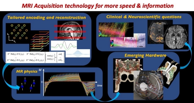
The research in our lab focuses on the development of new MRI acquisition technologies that can dramatically improve the speed, sensitivity and specificity of brain imaging. Our research explores approaches in designing tailored data acquisition & reconstruction algorithms using signal processing/optimization/ML methods, to take advantage of the underlying MR Physics and emerging hardware.
The goal is to create new imaging strategies that can help address important clinical & neuroscientific questions. The technologies that we have developed have enabled highly detailed brain data at unprecedented temporal and spatial resolutions, that have helped extract a wealth of quantitative information about brain structure and physiology. Some of these technologies have now been successfully translated as FDA-approved product, that are now being used daily in the clinic on the Siemens, GE and Phillips MRI scanners worldwide.
 – European Radiology
Clinical validation of Wave-CAIPI susceptibility-weighted imaging for routine brain MRI at 1.5 T
Objectives: Wave-CAIPI (Controlled Aliasing in Parallel Imaging) enables dramatic reduction in acquisition time of 3D MRI sequences such as 3D susceptibility-weighted imaging (SWI) but has not been clinically evaluated at 1.5 T. We sought to compare highly accelerated Wave-CAIPI SWI (Wave-SWI) with two alternative standard sequences, conventional three-dimensional SWI and two-dimensional T2*-weighted Gradient-Echo (T2*w-GRE), in patients undergoing routine brain MRI at 1.5 T.
 – Stroke
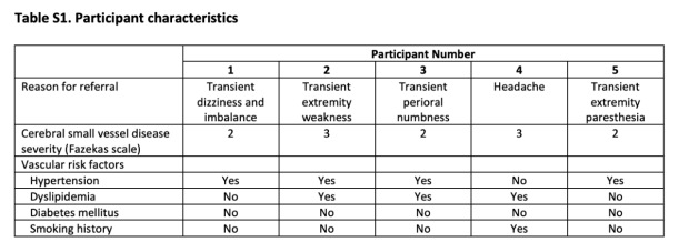
Detecting Silent Acute Microinfarcts in Cerebral Small Vessel Disease Using Submillimeter Diffusion-Weighted Magnetic Resonance Imaging: Preliminary Results
This research is part of a prospective observational study approved by the institutional review boards of the University Health Network and the University of Toronto. A board-certified neuroradiologist with 12 years of experience reviewed the submillimeter DWI, conventional DWI, and routine brain sequences side-by-side for each participant and identified acute and subacute microinfarcts.
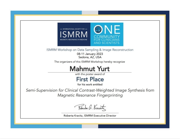
Title: Semi-Supervision for Clinical Contrast-Weighted Image Synthesis from Magnetic Resonance Fingerprinting. Summary of the abstract: Our work proposes a semi-supervised model for clinical contrast-weighted image synthesis from magnetic resonance fingerprinting with a training protocol based on highly accelerated acqusitions for more diverse data collection and reduced scan time.
- MIT EECS
Congratulation to Sid Iyer for his successful thesis defense!
Doctoral Thesis: On Improving the Acquisition and Reconstruction of Spatio-Temporal Magnetic Resonance Imaging
- Radiology
Congratulations to Congyu Liao and Nan Wang for being selected as 2022 ISMRM Junior Fellows!
Congyu Liao, PhD, an Instructor in Radiology, and Nan Wang, PhD, a Postdoctoral Scholar, both in the Setsompop Lab, were inducted as Junior Fellows. The ISMRM Junior Fellow Program was established to recognize outstanding researchers and clinicians at an early stage in their careers, with an established and long-term commitment to ISMRM.
- Radiology
Promotion to Instructor (faculty) to Congyu Liao!
Congratulations to Congyu Liao for his promotion to Instructor (faculty) in the department of Radiology, Stanford.
- ISMRM
2022 ISMRM AMPC - Congrats Uten Yarach!
Congratulations to Uten Yarach for being selected to join the Annual Meeting Program Commitee (AMPC) of the ISMRM!