Department Report Image Contest Winners
January 31, 2020
In preparing the 2019 Department Report, an "image contest" was held to collect creative and artistic images that comprehensively represented the breadth of research in radiology. The contest was open to all department members, and images had to be based on work performed while at Stanford.
Contest submissions included image representing the spectrum of research areas in the department–instrumentation-based images, molecular images results, AI-based renderings , and microscopy, to name a few. We sincerely thanks each researcher who took the time to prepare and submit an image for consideration, and for sharing their work wit us.
Our congratulations go to the researchers for their winning images and also to the Honorable Mention. All image, except for the First Prize image on the cover can be found on various pages throughout the Report. Thank you for participation and contributions to the Department Report.
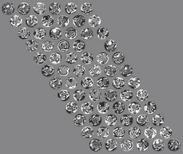
1st Place
Ningrui Li
PhD Student
1st Place - Ningui Li, PhD Student
Laboratory of Kim Butts Pauly, PhD, Radiological Sciences Laboratory
Shear waves were induced in a cylindrical gelatin phantom using a mechanical vibrator, causing them to scatter and reflect. Their displacements were imaged using a phase-contrast 3T MRI technique (MR elastography). This work highlights the importance of image reconstruction algorithms. Each individual wave image (represented by each circle) is chaotic and difficult to interpret by itself. However, thousands of wave images can be thoughtfully fused together using an image reconstruction algorithm to produce a single image representing the gelatin's mechanical properties. This is symbolized by the careful, structured positioning of the circular wave images into a coherent pattern, which contrasts with the disarray found within each individual wave image. This imaging technique can be used to help clinicians locate elusive tumors in cancer patients. Unfortunately, we are often times limited by the amount of information we can collect, as well as by invalid assumptions made by our algorithm. The void in this image, due to these limitations, symbolizes the gap in our knowledge of the underlying anatomical structures that we are trying to image.
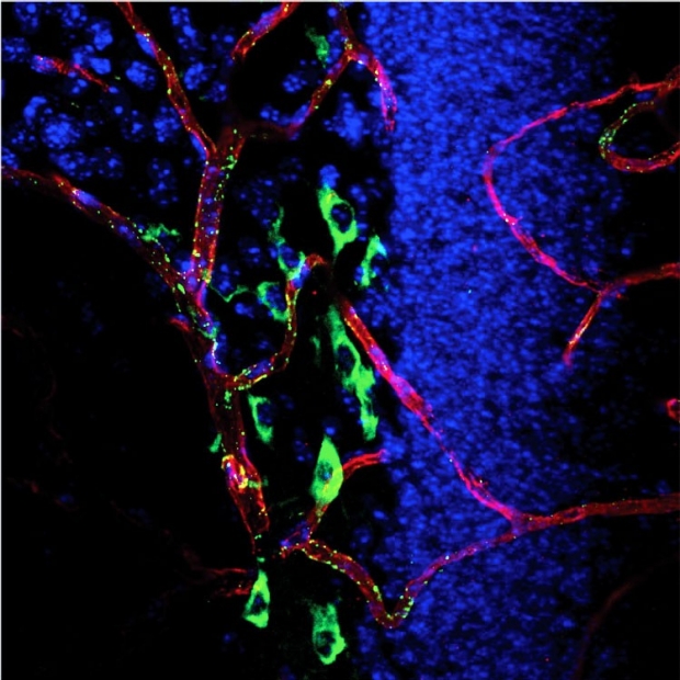
2nd Place
Marc Stevens, PhD
Postdoctoral Fellow
(page 25)
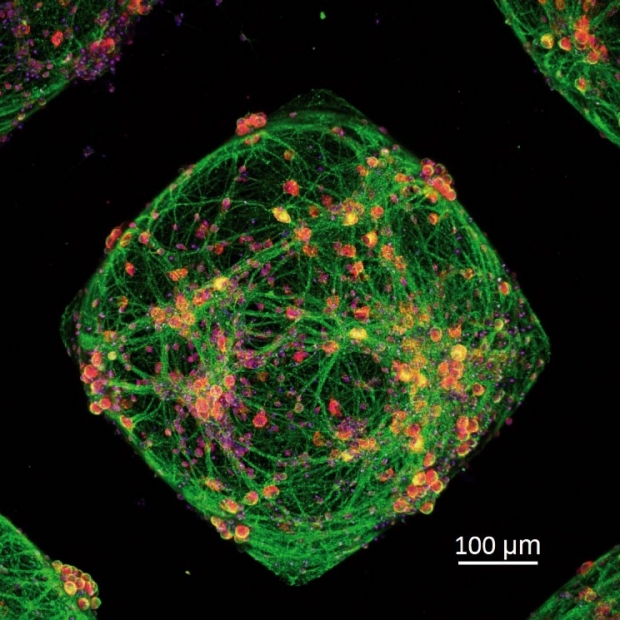
3rd Place
Tanchen Ren, PhD
Postdoctoral Scholar
(page 32)
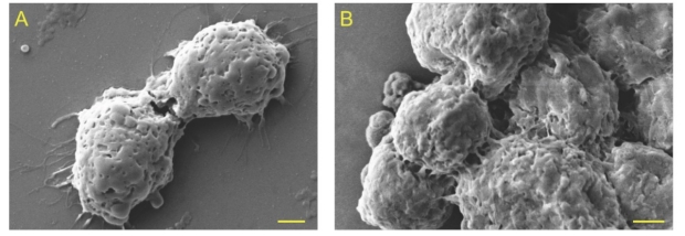
Honorable Mention
Jie Wang, PhD
Postdoctoral Scholar
(page 59)