Applying 3D Upper Airway Ultrasound Imaging to Clinical Diagnosis of Obstructive Sleep Apnea
To “see” the airway: how it collapses during snoring for our patients with obstructive sleep apnea remains a challenge. At Stanford, we started a study using ultrasound imaging to “see” your airway in a non-invasive manner without radiation.
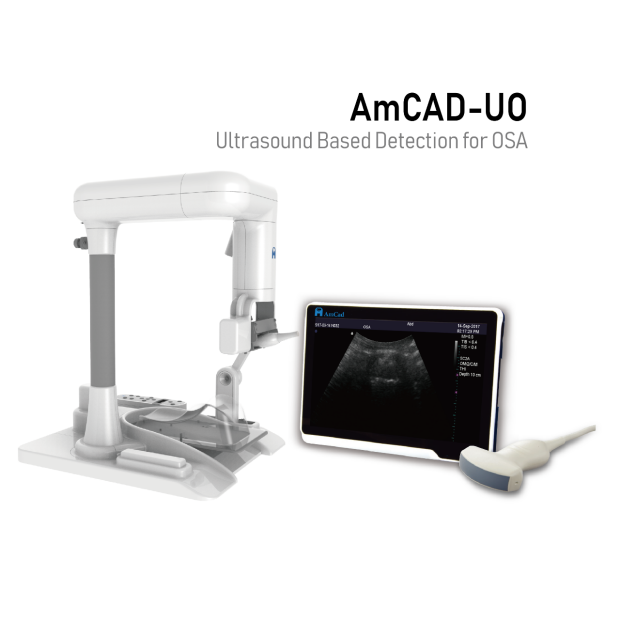
An FDA-approved ultrasound device build on artificial intelligence capability (AmCAD UO) is used to see your airway. It is then compared to traditional nasopharyngoscopy (i.e., a flexible tube is inserted into the nose to see the airway). Part of your routine clinic examination.
We hope that in the future, ultrasound visualization may become a better and easier way for you and your physician to see the airway affected by snoring and obstructive sleep apnea.
For participation in the study, the study coordinator would guide you through the following:
- If you agree to participate in the study, reviewing and signing the study consent form
- Instructing you on the four breathing modalities that are performed during the study
- Normal breathing
- Muller maneuver- a maneuver where you are asked to pinch your nostrils closed, and inhale through your nose
- Tongue rolling- a maneuver where you are asked to fold your tongue back
- Jaw protrusion- a maneuver where you are asked to move your lower jaw forward
The study takes approximately 20 minutes to complete and is done after one of your clinical appointments - there is no need for any additional visits.
If you are interested in participating, please contact the study coordinator, Vivian Liu at vivian44@stanford.edu or 650-721-7570.
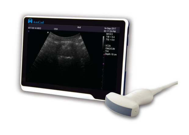

Artificial Intelligence to See the Airway Via Ultrasound
For any questions regarding Participants’ Rights, call 1-866-680-2906