
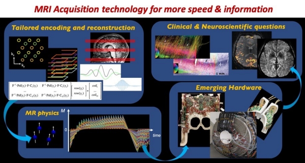
The research in our lab focuses on the development of new MRI acquisition technologies that can dramatically improve the speed, sensitivity and specificity of brain imaging. Our research explores approaches in designing tailored data acquisition & reconstruction algorithms using signal processing/optimization/ML methods, to take advantage of the underlying MR Physics and emerging hardware.
The goal is to create new imaging strategies that can help address important clinical & neuroscientific questions. The technologies that we have developed have enabled highly detailed brain data at unprecedented temporal and spatial resolutions, that have helped extract a wealth of quantitative information about brain structure and physiology. Some of these technologies have now been successfully translated as FDA-approved product, that are now being used daily in the clinic on the Siemens, GE and Phillips MRI scanners worldwide.
 – Wiley Online Library
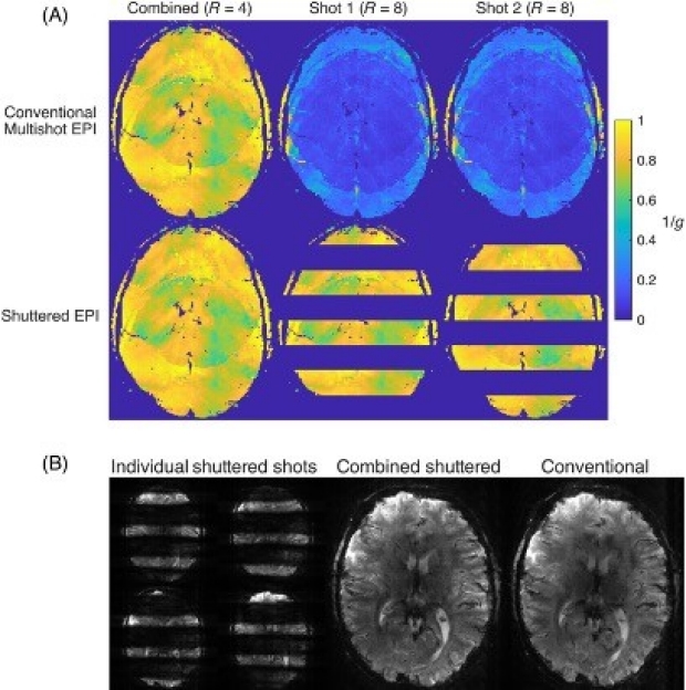
High‐resolution motion‐ and phase‐corrected functional MRI at 7 T using shuttered multishot echo‐planar imaging
Purpose: To achieve high-resolution multishot echo-planar imaging (EPI) for functional MRI (fMRI) with reduced sensitivity to in-plane motion and between-shot phase variations.
 – Nat Commun.
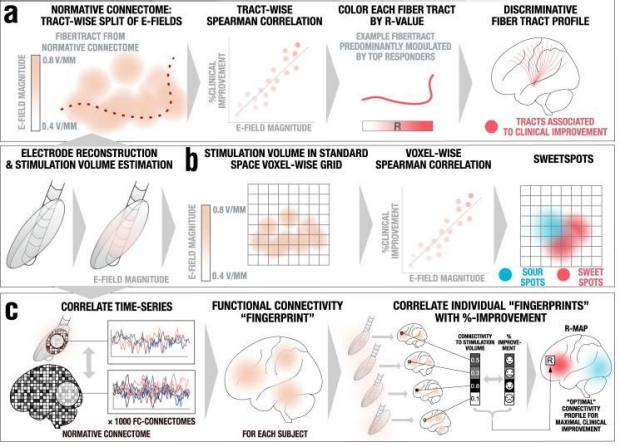
Optimal deep brain stimulation sites and networks for stimulation of the fornix in Alzheimer’s disease
Deep brain stimulation (DBS) to the fornix is an investigational treatment for patients with mild Alzheimer’s Disease. Outcomes from randomized clinical trials have shown that cognitive function improved in some patients but deteriorated in others.
 – Eur Radiol
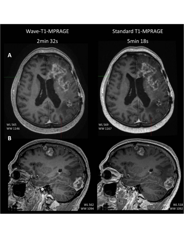
Validation of a highly accelerated post-contrast wave-controlled aliasing in parallel imaging (CAIPI) 3D-T1 MPRAGE compared to standard 3D-T1 MPRAGE for detection of intracranial enhancing lesions on 3-T MRI - European Radiology
Objectives: High-resolution post-contrast T1-weighted imaging is a workhorse sequence in the evaluation of neurological disorders. The T1-MPRAGE sequence has been widely adopted for the visualization of enhancing pathology in the brain. However, this three-dimensional (3D) acquisition is lengthy and prone to motion artifact, which often compromises diagnostic quality.
 – European Radiology
Clinical validation of Wave-CAIPI susceptibility-weighted imaging for routine brain MRI at 1.5 T
Objectives: Wave-CAIPI (Controlled Aliasing in Parallel Imaging) enables dramatic reduction in acquisition time of 3D MRI sequences such as 3D susceptibility-weighted imaging (SWI) but has not been clinically evaluated at 1.5 T. We sought to compare highly accelerated Wave-CAIPI SWI (Wave-SWI) with two alternative standard sequences, conventional three-dimensional SWI and two-dimensional T2*-weighted Gradient-Echo (T2*w-GRE), in patients undergoing routine brain MRI at 1.5 T.
The Academy for Radiology and Biomedical Imaging Research announces it 2023 Class of Distinguished Investigators. Established in 2012 to acknowledge and celebrate high levels of achievement in the field of imaging research. The award is presented at a ceremony held during the annual RSNA meeting. Recipients of the award will become members of the Council of Distinguished Investigators of the Academy.
- ISMRM
2023 ISMRM Magna Cum Laude Merit Award to Congyu Liao
"Flexible use of AC/DC coil for eddy-currents and concomitant fields mitigation with applications in diffusion-prepared non-Cartesian sampling" - Received Magna Cum Laude
- ISMRM
2023 ISMRM Magna Cum Laude Merit Award to Mahmut Yurt
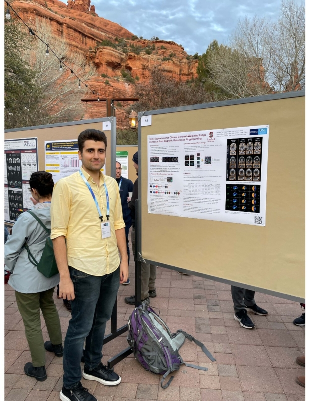
"Semi-Supervision for Clinical Contrast-Weighted Image Synthesis from Magnetic Resonance Fingerprinting" - Received Magna Cum Laude
- ISMRM
2023 ISMRM Summa Cum Laude Merit Award to Mahmut Yurt
"Conditional Denoising Diffusion Probabilistic Models for Inverse MR Image Recovery" - Received Summa Cum Laude & AMPC award, highlighted as one of the top 100 abstracts in ISMRM
- ISMRM
2023 ISMRM Summa Cum Laude Merit Award to Yannick Brackenier
“Towards rapid and accurate navigators for motion and B0 estimation using QUEEN (QUantitatively-Enhanced parameter Estimation from Navigators)” - Received Summa Cum Laude