
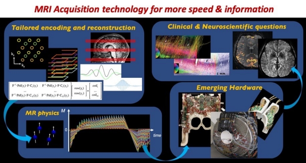
The research in our lab focuses on the development of new MRI acquisition technologies that can dramatically improve the speed, sensitivity and specificity of brain imaging. Our research explores approaches in designing tailored data acquisition & reconstruction algorithms using signal processing/optimization/ML methods, to take advantage of the underlying MR Physics and emerging hardware.
The goal is to create new imaging strategies that can help address important clinical & neuroscientific questions. The technologies that we have developed have enabled highly detailed brain data at unprecedented temporal and spatial resolutions, that have helped extract a wealth of quantitative information about brain structure and physiology. Some of these technologies have now been successfully translated as FDA-approved product, that are now being used daily in the clinic on the Siemens, GE and Phillips MRI scanners worldwide.
 – Nat Methods.
Next-generation MRI scanner designed for ultra-high-resolution human brain imaging at 7 Tesla
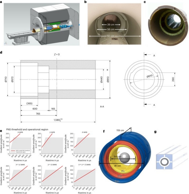
Abstract: To increase granularity in human neuroimaging science, we designed and built a next-generation 7 Tesla magnetic resonance imaging scanner to reach ultra-high resolution by implementing several advances in hardware. To improve spatial encoding and increase the image signal-to-noise ratio, we developed a head-only asymmetric gradient coil (200 mT m-1, 900 T m-1s-1) with an additional third layer of windings.
 – Magn Reson Med
DTI‐MR fingerprinting for rapid high‐resolution whole‐brain T1, T2, proton density, ADC, and fractional anisotropy mapping
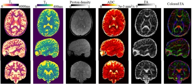
Purpose: This study aims to develop a high-efficiency and high-resolution 3D imaging approach for simultaneous mapping of multiple key tissue parameters for routine brain imaging, including T1, T2, proton density (PD), ADC, and fractional anisotropy (FA).
 – Magn Reson Med
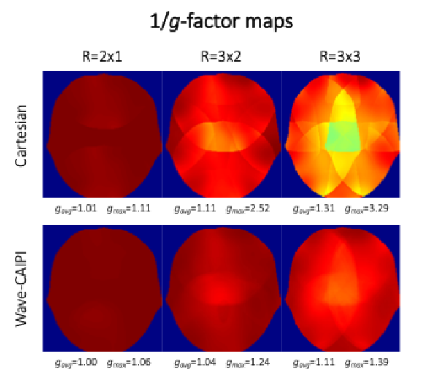
Time‐efficient, high‐resolution 3T whole‐brain relaxometry using 3D‐QALAS with wave‐CAIPI readouts
Purpose: Volumetric, high-resolution, quantitative mapping of brain-tissue relaxation properties is hindered by long acquisition times and SNR challenges. This study combines time-efficient wave-controlled aliasing in parallel imaging (wave-CAIPI) readouts with the 3D quantification using an interleaved Look-Locker acquisition sequence with a T2 preparation pulse (3D-QALAS), enabling full-brain quantitative T1 , T2 , and proton density (PD) maps at 1.15-mm3 isotropic voxels in 3 min.
 – NeuroImage
High-fidelity mesoscale in-vivo diffusion MRI through gSlider-BUDA and circular EPI with S-LORAKS reconstruction
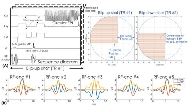
Purpose: To develop a high-fidelity diffusion MRI acquisition and reconstruction framework with reduced echo-train-length for less T2* image blurring compared to typical highly accelerated echo-planar imaging (EPI) acquisitions at sub-millimeter isotropic resolution.
The Academy for Radiology and Biomedical Imaging Research announces it 2023 Class of Distinguished Investigators. Established in 2012 to acknowledge and celebrate high levels of achievement in the field of imaging research. The award is presented at a ceremony held during the annual RSNA meeting. Recipients of the award will become members of the Council of Distinguished Investigators of the Academy.
- ISMRM
2023 ISMRM Magna Cum Laude Merit Award to Congyu Liao
"Flexible use of AC/DC coil for eddy-currents and concomitant fields mitigation with applications in diffusion-prepared non-Cartesian sampling" - Received Magna Cum Laude
- ISMRM
2023 ISMRM Magna Cum Laude Merit Award to Mahmut Yurt
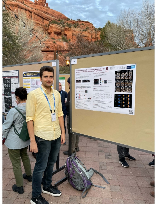
"Semi-Supervision for Clinical Contrast-Weighted Image Synthesis from Magnetic Resonance Fingerprinting" - Received Magna Cum Laude
- ISMRM
2023 ISMRM Summa Cum Laude Merit Award to Mahmut Yurt
"Conditional Denoising Diffusion Probabilistic Models for Inverse MR Image Recovery" - Received Summa Cum Laude & AMPC award, highlighted as one of the top 100 abstracts in ISMRM
- ISMRM
2023 ISMRM Summa Cum Laude Merit Award to Yannick Brackenier
“Towards rapid and accurate navigators for motion and B0 estimation using QUEEN (QUantitatively-Enhanced parameter Estimation from Navigators)” - Received Summa Cum Laude