Research Projects
Therapeutic Ultrasound
Ultrasound-based target-specific drug delivery
Ultrasound (US)-responsive drug-loaded nanoparticles are a promising solution for achieving target specificity by exposing them to ultrasound near tumors. We are currently investigating the efficient US exposure conditions for US-responsive nanoparticles and the therapeutic effectiveness of the nanoparticles (anticancer microRNAs, and chemicals) in large animal models, collaborating other research groups to enhance chemotherapy.
Fig. 1. In vivo delivery (canine model) by intravenous infusion of microbubbles and nanoparticles (PLGA-b-PEG) with an ultrasound transducer positioned on the tumor region (liver) for targeted microRNA ([1] S. U. Kumar et al., Adv. Ther., 2020).
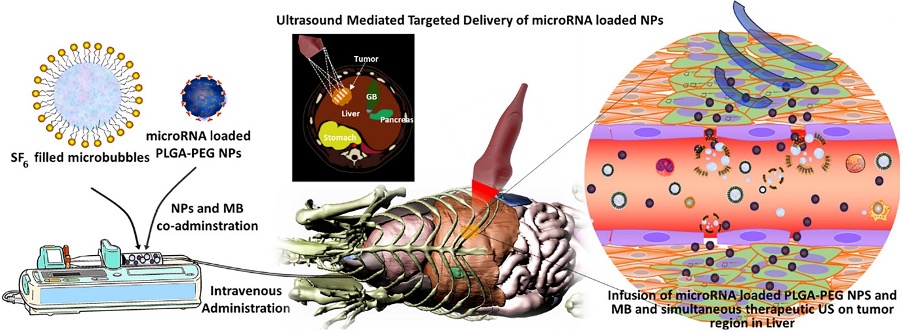
[1] S. U. Kumar, A. V. Telichko, H. Wang, D. Hyun, E. G. Johnson, M. S. Kent, R. B. Rebhun, J. J. Dahl, W. T. N. Culp and R. Paulmurugan, Acoustically Driven Microbubbles Enable Targeted Delivery of microRNA‐Loaded Nanoparticles to Spontaneous Hepatocellular Neoplasia in Canines. Advanced Therapeutics, 3(12), 2000120, 2020. doi: 10.1002/adtp.202000120
Passive cavitation mapping (PCM)
Precise and rapid cavitation monitoring is highly desirable for therapeutic ultrasound applications due to their safety. Passive cavitation monitoring approach which localizes acoustic emissions from cavitation using only receive beamforming, overcoming the limitation of active cavitation monitoring: time-consuming and non-real-time monitoring. Passive cavitation mapping (PCM), especially, enables spatial localization mapping of acoustic information of cavitation. We are currently developing and improving PCM algorithms to solve cavitation monitoring challenges in therapeutic ultrasound applications.
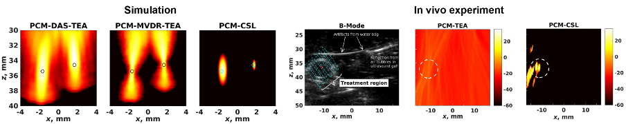
Fig. 2. Passive cavitation maps from the simulations with two sources placed at different locations shown on a 40-dB dynamic range (Left, [2] A. V. Telichko et al., IEEE Trans. Ultrason. Ferroelctr. Freq. Control, 2021).
Passive cavitation maps of in vivo mouse experiment. The tumor boundary is outlined with a white dashed line (Right, [3] A. V. Telichko et al., IEEE Trans. Ultrason. Ferroelctr. Freq. Control, 2021).
[2] A. V. Telichko, T. Lee, M. Jakovljevic and J. J. Dahl, Passive cavitation mapping by cavitation source localization from aperture-domain signals—Part I: Theory and validation through simulations. IEEE transactions on ultrasonics, ferroelectrics, and frequency control, 68(4), 1184-1197, 2021
[3] A. V. Telichko, T. Lee, D. Hyun, S. M. Chowdhury, S. Bachawal, C. D. Herickhoff, R. Paulmurugan and J. J. Dahl, Passive cavitation mapping by cavitation source localization from aperture-domain signals—Part II: Phantom and in vivo experiments. IEEE transactions on ultrasonics, ferroelectrics, and frequency control, 68(4), 1198-1212, 2021