Chronic Lung Disease of Infancy
Early contributions were focused upon the regulation of perinatal pulmonary vascular tone. Our Laboratory addressed the subcellular mechanisms that underlie the postnatal adaption of the pulmonary circulation. Key publications demonstrated that pulmonary artery endothelial cells produced endothelial-derived relaxing factor (EDRF), subsequently identified as nitric oxide (NO), in response to ventilation, oxygenation and shear stress. Further work demonstrated that oxygen, one of the key stimuli for perinatal pulmonary vasodilation, as well as NO acts via quantal and localized release of calcium from ryanodine-sensitive stores to prompt activation of the pulmonary artery smooth muscle cell calcium-sensitive potassium channels. These papers informed the development of several clinical trials wherein the efficacy of inhaled nitric oxide in persistent pulmonary hypertension of the newborn was established.
With improved obstetrical and neonatal care, more, smaller and less developmentally mature infants are surviving even extreme prematurity. Our Laboratory is interested in two fundamental questions surrounding chronic lung disease of infancy. First, whether and how might exposure of oxygen levels in excess of the normally low intrauterine oxygen tension state of the intrauterine environment might compromise lung development. Second, as pulmonary hypertension complicates chronic lung disease of infancy in ~30% of cases, our Laboratory is focusing on the molecular mechanisms that underlie the increase in pulmonary artery blood pressure and identification of novel therapeutic tools to control blood pressure and promote lung development.
Projects
The Loss of Pulmonary Pericytes is Associated with the Development of Bronchopulmonary Dysplasia
Researchers: Elizabeth A. Barnes, Xibing Che, Carsten Knutsen, Cristina M. Alvira, David N. Cornfield
Pulmonary pericytes are unique cells that offer support to and communicate with angiogenic endothelial cells. Using single cell RNA sequencing (scRNAseq) technique to determine differentially expressed genes in a mouse model of hyperoxia-induced lung injury to mimic bronchopulmonary dysplasia (BPD), pericytes were found to be nearly absent compared to normoxic mice by day 7 of life (P7). The identification of pericyte loss in a BPD mouse model is completely novel and suggests that the angiogenic deficiency found in patients with BPD may be pericyte dependent.
We have developed a novel protocol to isolate pericytes from various stages of pulmonary development. Preliminary results have shown that Hypoxia Inducible Factor (HIF) signaling may play a role in the early stages of hyperoxia-induced lung injury. Future studies will explore the role of pericytes in endothelial cell-driven pulmonary angiogenesis.
Hypoxia-Inducible Factor-1a in SM22a-Expressing Cells
Modulates Alveolarization
Researchers: Elizabeth A. Barnes, Carsten Knutsen, Alida Kindt, Xibing Che, Lihua Ying, Eloa Adams, Erika Gonzalez, Prajakta Oak, Anne Hilgendorff, Cristina M. Alvira, David N. Cornfield
In lung development, the most important process in alveolarization is when the alveolar ducts divide into alveolar sacs by secondary septation. We now know that angiogenesis drives this process. HIF-1a is a transcription factor important for angiogenesis, especially under conditions in which oxygen levels are limited. Infants born prematurely (less than 28 weeks gestational age) and treated with supplemental oxygen exhibit arrested lung development with impaired alveolarization and loss of angiogenesis, resulting in Bronchopulmonary Dysplasia (BPD).
The role of HIF-1a in the development of BPD remains unknown; therefore, to clarify the effects of HIF-1a, we created mice with a cell-specific deletion of HIF-1a. Mice with a SM22a cell-specific deletion of HIF-1a show impaired pulmonary development consistent with BPD. The loss of HIF-1a resulted in a loss of Angiopoietin 2, a secreted molecule essential for angiogenesis, suggesting that the loss of Angiopoietin 2 may be a factor in the development of BPD.
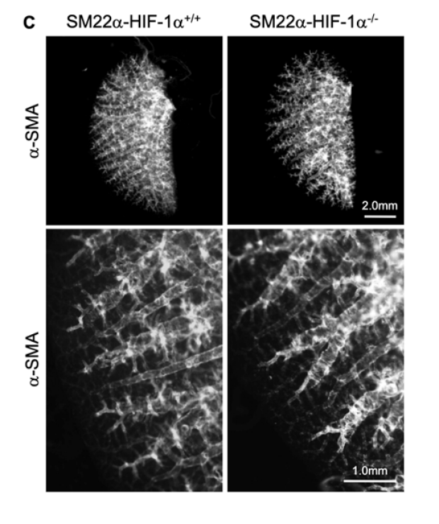
3-D images of a-SMA-stained vasculature from adult SM22a-HIF-1a mice. Left lobes were examined for the expression of a-SMA (white). Upper panels, scale bar 2.0mm. Lower panels, enlarged images, scale bar 1.0mm.
Stabilization of HIF-1α in SM22α-expressing Cells Prevents the Development of BPD in a Mouse Model of Lung Development
Researchers: Reiji Ito, Elizabeth A. Barnes, Xibing Che, Cristina M. Alvira, David N. Cornfield
Though the survival rates for preterm infants are improving, the incidence of chronic lung disease of infancy, or Bronchopulmonary Dysplasia (BPD), remains high. BPD is characterized by larger and fewer alveoli (air sacs). We have previously shown that the loss of HIF-1α expression in a mouse model of BPD impairs alveolarization and angiogenesis. Therefore, we sought to determine if HIF-1α stabilization in SM22α-expressing cells can limit hyperoxia-induced neonatal lung injury, a mouse model of BPD. In mice expressing stabilized HIF-1a, (SM22α-PHD1/2-/- mice) we found that stabilization of HIF-1α in pulmonary SM22α-expressing cells protects neonatal lung development despite prolonged hyperoxia exposure. In addition, Angiopoietin 2, microvascularization, and angiogenesis were increased in mice expressing stabilized HIF-1α.
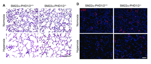
SM22a cell specific HIF-1a stabilization mitigates the effects of hyperoxia-induced neonatal lung injury. (A) H&E stained pulmonary tissues from postnatal day 14 (P14) SM22a-PHD1/2 mice neonatally exposed to normoxia or hyperoxia for 14 days. Scale bar 100microns. SM22a-PHD1/2-/- mice exhibit smaller alveoli with exposure to hyperoxia, indicating less lung injury compared to control mice. (D) Von Willebrand (red) stained pulmonary tissues from P14 SM22a-PHD1/2 mice neonatally exposed to normoxia or hyperoxia for 14 days. Scale bar 100 microns. SM22a-PHD1/2-/- mice exhibit an increase in von Willebrand stained vessels under both normoxic and hyperoxic conditions, indicating an increase in pulmonary angiogenesis compared to control mice.
Predisposition to Pulmonary Hypertension Linked to Hyperoxia-Induced Neonatal Lung Injury
Researchers: Reiji Ito, Elizabeth A. Barnes, David N. Cornfield
In Bronchopulmonary Dysplasia (BPD), lung function continues to deteriorate until adulthood leading to obstructive pulmonary disease. Moreover, ~20% of BPD patients suffer from Pulmonary Hypertension (PH), and in BPD patients diagnosed with PH there is a higher mortality rate (~50%).
In our SM22a-PHD1/2 BPD mouse model we have found that not only are SM22a-PHD1/2-/- mice protected from developing BPD, but as adults are protected from developing PH. These mice exhibit diminished pulmonary arterial wall thickness and right ventricular hypertrophy compared to control mice.
The SM22a-HIF-1a Mouse as a Model for Bronchopulmonary Dysplasia
Researchers: Elizabeth A. Barnes, Chihhsin Chen, Xibing Che, Ross Metzger, Cristina Alvira, David N. Cornfield
Bronchopulmonary dysplasia (BPD) affects infants born prematurely due to the lack of mature lung function. HIF-1a has been found to be an invaluable component in the maturing lung; therefore, we made a mouse lacking SMC-HIF-1a and found that these mice maintain an immature lung phenotype, resembling the human condition.
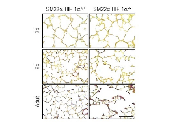
Mice without SMC-HIF-1a display disorganized elastin (brown) expression (right panels), consistent with BPD. Magnification 200x, calibration bar 200mm. 3d, 3 day old mice; 8d, 8 day old mice; Adult, adult mice.
People

Cristina Alvira, M.D.

Elizabeth A. Barnes, Ph.D.

Xibing Che, Ph.D.

Chihhsin Chen. M.S.

David N. Cornfield, M.D.

Bereketeab Haileselassie, M.D.

Reiji Ito, M.D., Ph.D.

Trang Dinh, Ph.D.