Molecular Regulation of Pulmonary Vascular Tone
Over the past several years, our lab has been engaged in experiments that address the regulation of pulmonary vascular tone. We have investigated the signal transduction pathway of molecules that play an important role in determining pulmonary vascular tone. Relatively recently, we identified a novel, and controversial, role for HIF-1a in the regulation of tone in lung via effects on myosin light chain phosphorylation in the pulmonary artery smooth muscle cells and in regulating expression of the b1 subunit of the calcium sensitive potassium channel. Further, we outlined a novel role for endothelin derived from PASMC in modulating the pulmonary vascular response to hypoxia. Most recently, we have undertaken a line of research using human tissue to ensure fidelity between findings in mouse models and human biology.
Projects
Characterization of the SM22a-PHD1/2 Mouse to Further Elucidate the Role of SM22a-HIF-1a in a Mouse Model of Hypoxia-induced Pulmonary Hypertension
Researchers: Elizabeth A. Barnes, Reiji Ito, Xibing Che, Chihhsin Chen, Sushma Reddy, and David N. Cornfield
In addition to the loss of PHD1 and PHD2 being protective in the heart, the loss of PHD1 and PHD2 in the lungs is protective regarding the development of PH in a hypoxia-induced mouse model. This is likely due to an increase in HIF-1a activity promoting angiogenesis.
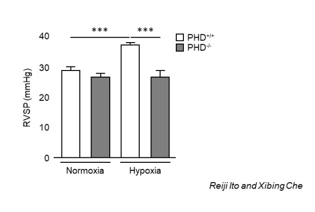
Pulmonary arterial pressures are increased in WT mice, but not in mice devoid of SMC-PHD1 and PHD2 under chronic hypoxic conditions.
Loss of PHD1/2 in SM22a-Expressing Cells Prevents Hypoxia-Induced Pulmonary Hypertension
Researchers: Elizabeth A. Barnes, Reiji Ito, Xibing Che, Cristina M. Alvira, David N. Cornfield
Pulmonary arterial hypertension (PAH) is a disease characterized by increased vasoconstriction and vascular remodeling. Pulmonary artery smooth muscle cells (PASMC) highly express the transcription factor HIF-1a, yet the role of HIF-1a in the development of PAH remains controversial. To address this issue, we generated a mouse expressing stabilized HIF-1a inSM22a-expressing cells (SM22a-PHD1/2). SM22a-PHD1/2-/- mice were protected from increases in right ventricular systolic pressures (RVSP), vasoconstriction, and vascular remodeling when exposed to hypoxia. These findings suggest that modulation of HIF-1a in SM22a-expressing cells may represent a therapeutic target for PAH.
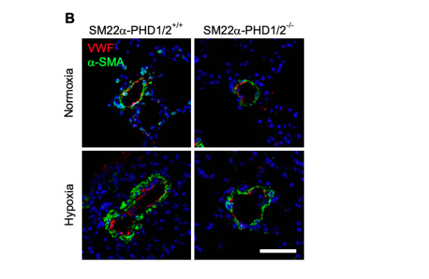
Vascular remodeling is attenuated in SM22a-PHD1/2-/- mice exposed to hypoxia. Assessment of arteriole muscularization in SM22a-PHD1/2 mice under normoxic and chronic hypoxic conditions. VWF-positive (red),a-SMA-positive (green) arterioles. 200x magnification, calibration bar 50microns.
Loss of Smooth Muscle Cell Hypoxia Inducible Factor-1a Underlies Increased Vascular Contractility in Pulmonary Hypertension
Researchers: Elizabeth A. Barnes, Chih-Hsin Chen, Oshra Sedan, David N. Cornfield
Pulmonary arterial hypertension (PAH) is an often fatal disease with limited treatment options. Pulmonary artery smooth muscle cells (PASMC) highly express the transcription factor HIF-1a, yet the role of HIF-1a in the development of PAH remains controversial. This study investigates the hypothesis that in patients with PAH, decreases in PASMC HIF-1a expression and activity underlie augmented pulmonary vascular contractility.
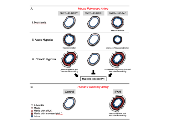
Role for PASMC HIF-1a in limiting vasoconstriction and vascular remodeling. (A) In SM22a-genetically altered mouse pulmonary arteries: the level of HIF-1a expression inversely correlates with pMLC expression, contraction, and vascular remodeling. (B) In human PAH patients, the level of HIF-1a inversely correlates with pMLC expression, contraction, and vascular remodeling.
Smooth Muscle Cells from Pulmonary Hypertensive Patients Secrete BMP2 to Induce Endothelial Cell Aberrant Angiogenesis
Researchers: Elizabeth A. Barnes, David N. Cornfield
It has been demonstrated that pulmonary artery endothelial cells (PAEC) that are devoid of functional BMPR2 become migratory when stimulated with BMP2, the ligand for BMPR2. We have found that SMC from patients with PH secrete BMP2 and promote PAEC dysfunctional migration. This event plays a key role in the pathogenesis of PH.
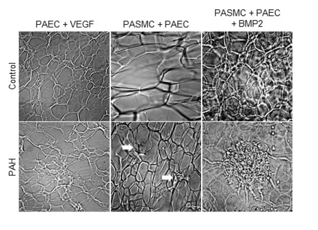
BMP2-induced angiogenesis. PAEC and PASMC from PH patients display dysfunctional angiogenesis in the presence of BMP2 (lower middle panel). Arrows represent small disorganized vascular masses. With the addition of exogenous BMP2, PAEC and PASMC from PH patients display further dysfunctional angiogenesis (lower right panel). Magnification 200x.
Metabolism of Pulmonary Artery Smooth Muscle Cells from Patients with Pulmonary Hypertension
Researchers: Elizabeth A. Barnes, Riddhita Mukherjee, Bereketeab Haileselassie, David N. Cornfield
Controversy exists regarding the metabolic state of smooth muscle cells (SMC) from pulmonary hypertensive (PH) patients. Therefore, we have examined SMC from PH patients in comparison to SMC. Preliminary results show that SMC from PH patients differ greatly compared to controls in that they are more adaptable to various cellular environments.
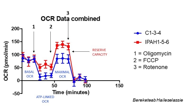
Oxygen consumption rates (OCR) of control (C1, C3, C4) and PH (IPAH1, IPAH 5, IPAH6) SMC. PH SMC exhibit enhanced ATP synthesis under conditions of cellular stress. PH SMC metabolic adaptability may play a role in the pathogenesis of PH.
The SM22a-PHD1/2-/- Heart
Researchers: Elizabeth A. Barnes, David N. Cornfield
The loss of PHD1 and PHD2 (prolyl hydroxylase domain) proteins in the mouse heart leads to an increase in cardiac microvessels. PHD proteins degrade HIF-1a, a major component of angiogenesis. Without SMC-PHD1 and PHD2 present, HIF-1a remains stable and is transcriptionally active. The increase in microvessels likely plays a protective role in the heart.
PHD1/2+/+ (wild-type, WT) Heart (right panel) and PHD1/2-/- Heart (left panel). Only the PHD1/2-/- heart remains functional ex vivo. Hearts from SM22a-PHD1/2 mice were examined ex vivo after Langendorff perfusion. Hearts from PHD1/2-/- mice with stabilized HIF-1a remained beating ex vivo for several minutes.
People

Elizabeth A. Barnes, Ph.D.

Chihhsin Chen, M.S.

Xibing Che, Ph.D.

David N. Cornfield, M.D.

Reiji Ito, M.D., Ph.D.