Graphics
Visual rendering is an essential aspect of a surgical simulation experience. The surgeon needs to be able to readily differentiate between tissue types to recognize critical structures and surgical landmarks.
In CardinalSim, graphic rendering is performed directly from the clinical volumetric imaging data. The system employs modern programmable graphics processors (GPUs) using a technique known as GPU-accelerated ray casting. This approach allows real-time rending of sufficiently realistic images even using relatively inexpensive commodity computing hardware. The maintenance of a volumetric representation of structures preserves the potential to capture subtle translucency effects critical for surgeons to identify structures through thin volumes of bone.
Enter Heading TextSchematic representation of visual rendering using GPU-accelerated ray casting
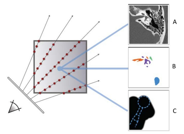
Rays are simultaneously cast through the following:
- (A) The primary volumetric dataset (usually CT)
- (B) Label field volumes indicating which voxels are part of which structures
- (C) Mask volumes, which indicate what portion of bone has been surgically removed
Imaging datasets can be assigned transfer functions to map material properties to the grayscale values of individual voxels
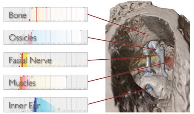
- Each volumetric labelfield derived during segmentation can be assigned a unique transfer function.
- This image shows the assignment of 5 discreet transfer functions to differentiate the structures in this microCT scan of a human cadaveric temporal bone core (temporal bone, ossicles, facial nerve, muscles, and inner ear).
Video capture of a transfer function being applied to a clinical sinus CT. Color and opacity may be adjusted interactively
Video capture of a right temporal bone scan obtained from a clinical scan of a patient undergoing cochlear implantation
- CardinalSim offers the ability to see the primary data from the underlying CT scan in an interactive tri-planar view to help maintain orientation of the 3D reconstruction to more conventional planar projections.
Visual representations generated during simulated surgical rehearsal can replicate critical anatomic findings seen at actual surgery
- Image (A) shows the intraoperative exposure of a cholesteatoma as seen during a left tympanomastoidectomy.
- Image (B) shows the simulated representation of that same patient’s anatomy in CardinalSim. The cholesteatoma (yellow arrow) was segmented from a fused diffusion-weighted MRI sequence.
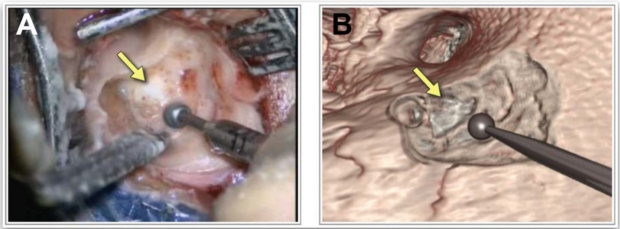
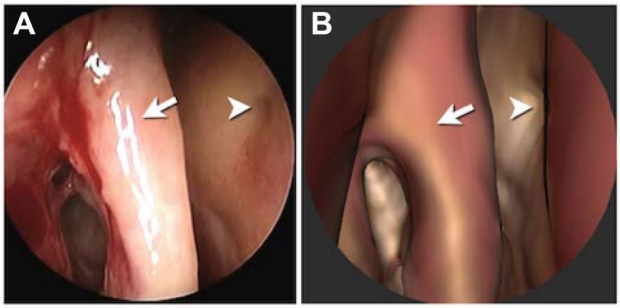
These images are from a patient undergoing resection of a tumor from the right middle ear. The tumor (arrowhead) extends under the long process of the malleus (arrow)
- Image (A) shows the intraoperative view.
- Image (B) shows the simulated representation of the same patient’s anatomy in CardinalSim.
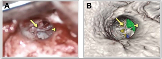
Endoscopic images from a patient undergoing endoscopic sinus surgery. Features of the septum (arrowhead) and attachment of the middle turnbinate (arrow) are shown
- Image (A) shows the intraoperative view.
- Image (B) shows the simulated representation of the same patient’s anatomy in CardinalSim.