Validation Studies
Endoscopic Sinus Surgery
Endoscopic sinus surgery (ESS) is a technically challenging endeavor that will benefit if a surgeon can explore patient-specific anatomy prior to undertaking actual surgery. Important structures such as the orbit, optic nerve, carotid artery, cranial vault, and nasolacrimal system are at risk for injury, and can be considerably altered by the presence of disease.
A thorough understanding of the anatomic variability encountered during surgery is critical in achieving successful outcomes and decreasing complications.

Angles of surgical approach and regions of limited access can be difficult to envision by examining a series of standard two-dimensional radiographic slices. Currently there is no intuitive method for the realistic interaction with preoperative imaging data.
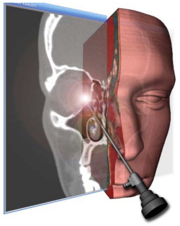
CardinalSim offers new technology to allow surgeons to view, manipulate, and even feel a patient’s anatomy based on preoperative imaging, which could provide considerable improvement over traditional planning methods.

The 3-dimensional virtual surgical environment may allow for enhanced pre-operative planning and rehearsal, with the goal of improving patient outcomes, decreasing complication rates, and enhancing technical skills.
We undertook a study to assess the design, features, and initial experience of CardinalSim for rhinologic procedures. The system allows a surgeon to interact with patient-specific, 3-dimensional reconstructions of sinus CT datasets using a modified haptic (touch feedback) interface device driving a virtual endoscope.
Initial evaluation of the simulation system was performed using CT datasets from patients who underwent endoscopic sinus surgery at the Stanford Sinus Center. The patients’ surgical procedures were recorded to enable comparisons between the virtual endoscopic views and actual intraoperative endoscopic views.
Using the intra-operative video as a reference, the virtual endoscope was navigated through the nasal cavity to the site of the procedure. Images of the virtual procedure were captured during this process to facilitate comparison. Anatomical correlations, key structural features, and other similarities between the intra-operative images and the simulated procedure were noted.
CardinalSim allowed for high fidelity anatomic correlation between virtual and intraoperative findings based on both the surgeons’ subjective impression, and the comparison of virtual and intraoperative images.
The graphics rendering frame rate approximates that of smooth endoscopic video.

A key element in the design of the system is its capability to directly load pre-operative CT DICOM image series.
CardinalSim offers the ability to interact with a virtual patient through a simulated endoscope similar to that used in ESS. Patient-specific surgical simulation offers a new frontier in patient care and medical education for this common procedure.
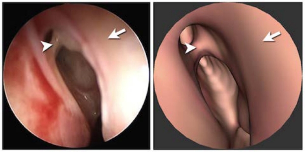
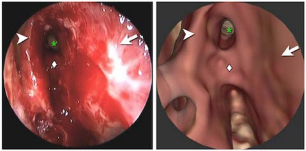
Intraoperative ESS images are shown on the left, with corresponding virtual sugical images from CardinalSim shown on the right.
The images show a high degree of accuracy in reproducing surgically-relevant anatomic detail in the nasal cavity and sinuses.
Reference:
Parikh, S. S. et al. Integration of patient-specific paranasal sinus CT data into a virtual surgical environment. American Journal of Rhinology (2009).
Temporal Bone Surgical Replication
To benefit preparation for otologic surgical procedures, a rehearsal system must allow relatively inexperienced users to reliably replicate key steps in actual ear procedures.
We undertook to assess this by asking a group of residents to replicate steps in a variety of otologic procedures.
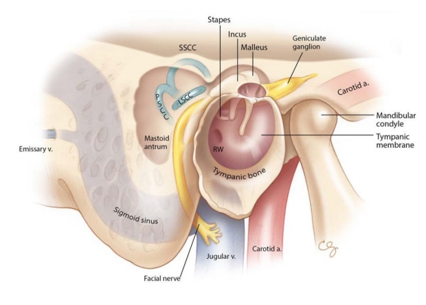
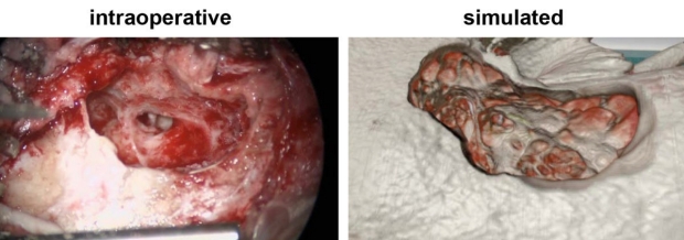
These images are from a patient undergoing right cochlear implantation. Mastoidectomy has been performed, and the facial recess opened.
Close-up of the completed facial recess approach demonstrating the stapes superstructure (arrow) and round window niche (arrowhead).
These images demonstrate the ability of surgeons to replicate the critical anatomic structures and relationships related to tympanomastoid surgery.
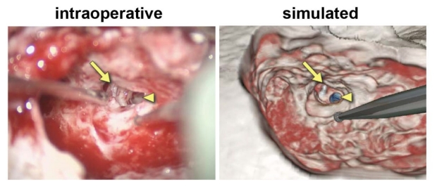
Intraoperative and simulated exposure of a middle ear adenoma. These images are from a patient with a middle ear adenoma in the hypotympanum. The adenoma (arrowhead) can be seen extending superiorly under the long process of the malleus (arrow).
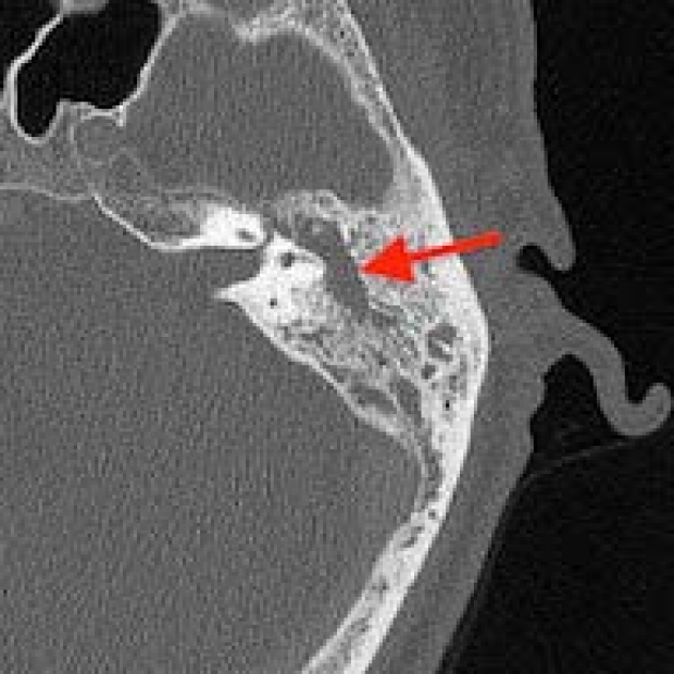
Real and simulated middle fossa exposure of a superior semicircular canal dehiscence (arrow).


Residents using CardinalSim to replicate intraoperative findings found it a useful tool for better understanding surgical anatomy.
The haptics, stereo graphics, and user interface were all found to add to the experience of using the system.
In a related study, fused CT and MRI sequences from patients with cholesteatoma were imported into CardinalSim to replicate tympanomastoid procedured.
The fused datasets showing both cholesteatoma and bony anatomy were used as the basis for simulation.

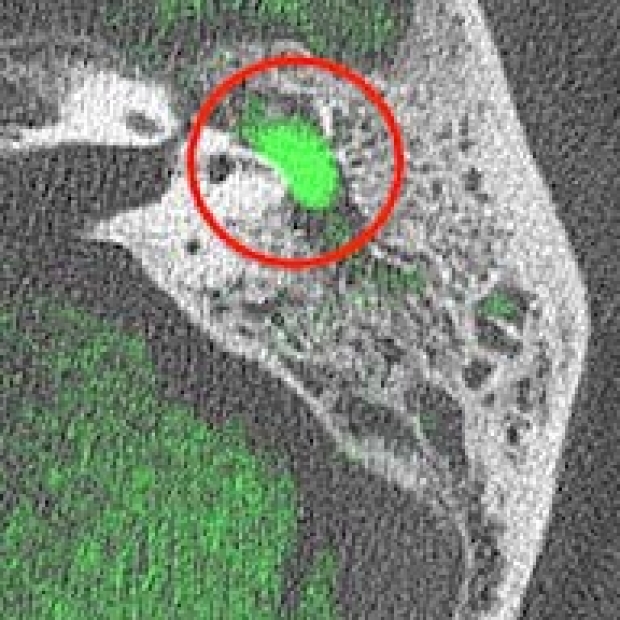
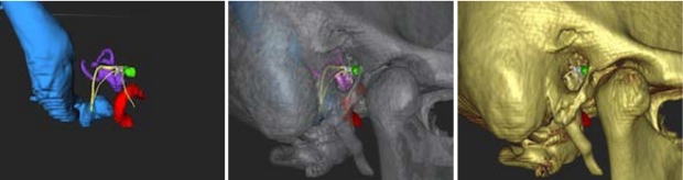
Models were constructed from segmented fused datasets.

Simulated dissection of the fused model demonstrated realistic relationship of the cholesteatoma (arrow) to the surrounding mastoid bone.