Bio
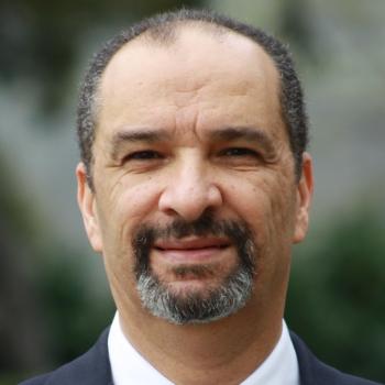
Tarik Massoud is a Professor of Neuroradiology and Molecular Imaging in the Department of Radiology, Stanford University School of Medicine, where he directs LEMNI (the Laboratory of Experimental and Molecular Neuroimaging), and is an Attending Neuroradiologist in Stanford Health Care. He qualified from the Medical School of the Royal College of Surgeons in Ireland and then served as intern to two inspirational medical giants of their days, Dr. William H. (Willie) Bisset at the Royal Hospital for Sick Children in Edinburgh, UK, and Professor Sir Raymond (Bill) Hoffenberg, PRCP, at the Queen Elizabeth Hospital in Birmingham, UK. He trained in Radiology and Neuroradiology in Oxford, UCLA, and the University of Michigan, and is a Fellow of the Royal College of Radiologists in London. He holds a research MD degree (NUI) in experimental neuroimaging (work conducted at UCLA), and a University of Cambridge PhD in molecular imaging (work conducted at the Crump Institute for Molecular Imaging at UCLA, and the Molecular Imaging Program at Stanford, Gambhir laboratory). From 2000 to 2013 he was a University Lecturer and Honorary Consultant in Neuroradiology at the University of Cambridge School of Clinical Medicine and Addenbrooke’s Hospital in Cambridge, UK. He was formerly an Assistant and Associate Professor of Radiology at UCLA, and held visiting Associate Professorships at Columbia, New York, and MCW, Milwaukee. He has published extensively and won numerous awards at scientific meetings. His papers in experimental interventional neuroradiology and molecular imaging are widely cited. He has been a peer reviewer for dozens of international medical journals, as well as other medical charities and governmental funding agencies. Until 2023 he was founding Editor-in-Chief of the journal Reports in Medical Imaging, and is an editorial board member for numerous biomedical journals. He is the senior author or editor of nine books, including "Glioblastoma: State-of-the-Art Clinical Neuroimaging", "Basilar Artery: A Clinical Review", "Glioblastoma Resistance to Chemotherapy: Molecular Mechanisms and Innovative Reversal Strategies", "Neuroimaging Anatomy: Parts 1 and 2", and "What Radiology Residents Need to Know: Neuroradiology". In 2016 he was awarded a Special Faculty Permit ('eminent physician license') by the Medical Board of the state of California. In 2022, he was honored with a Lifetime Achievement Award by the Royal College of Surgeons in Ireland School of Medicine.


