Improved Arterial Spin Labeling (ASL) Perfusion Imaging
Ferumoxytol (Feraheme, AMAG Pharmaceuticals Inc., Cambridge, MA) is an Ultrasmall SuperParamagnetic Iron Oxide particle (USPIO) compound recently approved for human use as a treatment for iron-deficiency anemia. Ferumoxytol can also be used off label as a strong T2* MR contrast agent, which could open many new possibilities in clinical MR imaging, including high-resolution perfusion mapping, enhanced fMRI activation detection, or imaging of inflammation processes in various diseases.
Steady-State and Dynamic perfusion Mapping
(T. Christen, W. Ni)
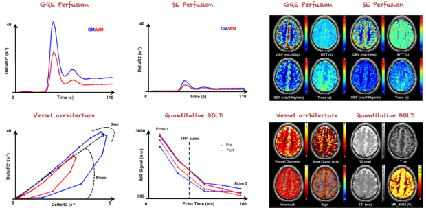
It has been recently suggested that following a bolus of contrast agent (CA) with a combined spin-echo (SE) and gradient-echo (GRE) sequence can yield valuable information about vessel architecture and oxygenation [1-2]. The pairwise R2* and R2 data points create a vortex curve whose characteristics can describe the effect of antiangiogenic treatments in patients with brain tumors [1]. More importantly, the method can identify patients who would benefit from therapies. While promising, the approach might, however, suffer from problems related to low signal-to-noise ratio and CA leakage if a gadolinium based CA is used. As an alternative, we propose in this project to follow an injection of ferumoxytol, which creates large magnetic susceptibility effects and whose large size confines it intravascularly. We study the feasibility of the vortex curve approach with USPIO icontrast and compare the results with bolus perfusion and quantitative BOLD (qBOLD) analyses.
The long half-life of ferumoxytol in the blood also enables alternative approaches to Dynamic susceptibility Contrast (DSC) for perfusion imaging. The steady-state susceptibility contrast method [3] that consists of acquisitions of T2* or T2 maps before and after contrast agent injection has previously been limited to use in animals. We have shown that quantitative high-resolution Cerebral Blood Volume maps (1mm istotropic spatial resolution) can be obtained in the human brain using this approach [4]. This should enable the evaluation of small lesions and allow comparison of intra and inter-patients blood volume changes during treatment or challenges.
Enhanced sensitivity for functional MRI
(D. Qiu, T. Christen)
Functional MRI (fMRI) brain studies performed in the presence of a steady-state or “blood pool” contrast agent yields activation maps that are weighted for cerebral blood volume (CBV). Previous animal experiments suggested significant contrast-to-noise ratio (CNR) improvements, but these studies had not yet been performed in humans due to the lack of availability of a suitable agent. We have reported the use of the ferumoxytol for functional brain activation in humans, termed contrast enhanced functional blood volume imaging (CE-fBVI) [5].
Four subjects were scanned during a unilateral finger tapping task with standard blood–oxygen level dependent (BOLD) imaging before contrast and CE-fBVI after contrast injection. The CE-fBVI response showed both a fast (5.8±1.3 s) and a slow (75.3±27.5 s) component of CBV response to stimuli. A significant CNR gain of approximately 2–3 was found for CE-fBVI compared to BOLD fMRI. Interestingly, less susceptibility-related signal dropouts were observed in the inferior frontal and temporal lobes with CE-fBVI. The combination of higher CNR and better spatial specificity, enabled by CE-fBVI using blood pool USPIO contrast agent opens the door to higher resolution brain mapping.

References: [1] Emblem et al, Nature Med, 2013. [2] Xu et al, Magn Reson Med, 2011. [3] Troprès et al, Magn Reson Med, 2001. [4] Christen et al, Magn Reson Med, 2012. [5] Qiu et al, NeuroImage, 2012.