Quantitative Susceptibility Mapping (QSM)
Magnetic Susceptibility at Ultra High Field (7T)
(J.M Park)
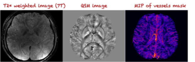
Ultra high field MR allows us to acquire images with excellent T2*-weighted contrasts in a sub-mm spatial resolution. After QSM analysis, we are able to differentiate subtle changes in magnetic susceptibility that permit scientific and clinical investigations of brain structures with superb conspicuity and delineation (e.g., sub-thalamic regions). High resolution venograms can also be created using the great contrast existing between blood vessels and surrounding tissues.
Plural Contrast Imaging
(S. Holdsworth, S. Soman)
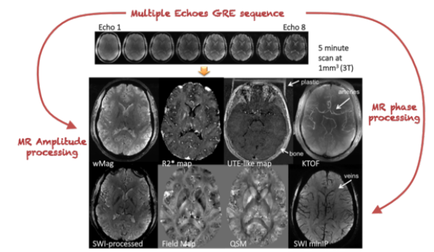
In previous work, we have extensively used the 3D multi-echo Gradient-Recalled Echo (ME-GRE) sequence coupled with our in-house built post-processing software (SILC) [1] for high-resolution Quantitative Susceptibility Mapping (QSM) [2-5]. We are currently using this technique in studies involving the use of the iron-oxide contrast-agent feraheme in brain tumor, stroke, and inflammatory nerve root imaging.
Previous work has shown that the 3D ME-GRE technique allows for the simultaneous generation of naturally co-registered images with various contrasts, including T2*, T1, phase, Susceptibility-Weighted Imaging (SWI), FLAIR T2* and Time-of-Flight (ToF)[6-7]. We are expanding upon this work with a more comprehensive plural-contrast approach, by implementing an advanced in-house built post-processing toolkit that also includes QSM, p-space [8], myelin-water fraction, ultra-short TE-like contrast, and vessel oxygenation.Some of these contrast mechanisms are shown in Fig. 1. We hypothesize that the information obtained from these multiple contrast mechanisms reveal complimentary image features, could improve our understanding of both normal tissue anatomy as well as changes in tissue in various pathological conditions, and potentially replace the need for multiple image sequences in clinical practice.
Magnetic Susceptibility in Pediatric Tumor Patients
(S. Holdsworth, J.M. Park, S. Soman)
Magnetic susceptibility measures various metabolic characteristics such as iron contents and oxygenation, providing useful tissue contrasts. While most previous studies have focused on measurements in healthy tissues, we are currently evaluating the QSM technique in pediatric oncological studies, facing the challenges of reliable susceptibility measurements in massive hemorrhagic regions and calcification of tumor.

References: [1] Qiu D et.al. Intl Soc Magn Reson Med. 2010; 4470. [2] Schweser F et.al. Neuroimage. 2011;54(4):2789–807. [3] Salomir R et.al. Concepts in Magnetic Resonance Part B: Magnetic Resonance Engineering. 2003;19B(1):26–34. [4] Marques JP, Bowtell R. Concepts in Magnetic Resonance Part B: Magnetic Resonance Engineering. 2005;25B(1):65–78. [5] Liu T et al. NMR Biomed. 2011;24(9):1129–36. [6] J. Luo et.al. NeuroImage 60: 1073–1082 (2012). [7] A. Deistung et.al. JMRI 29:1478–1484 (2009). [8] C. Liu, W. Li. NeuroImage 67: 193–202 (2013).