Descemet Endothelial Thickness Comparison Trial I
Descemet Endothelial Thickness Comparison Trial (DETECT) I is a multi-center, outcome assessor-masked, placebo-controlled clinical trial randomizing 160 patients in a 2x2 factorial design. The purpose of this study is to determine differences in visual outcomes between two types of corneal transplant surgeries, ultrathin Descemet stripping automated endothelial keratoplasty (UT-DSAEK) and Descemet membrane endothelial keratoplasty (DMEK), and to determine the effect of rho-kinase inhibitors on endothelial cell loss.
Stanford is currently accepting patients for this trial.
Stanford Investigator(s):
Intervention(s):
- drug: Topical Ripasudil
- drug: Topical Placebo
Eligibility
Inclusion Criteria:
- Dysfunctional endothelium from Fuchs Endothelial Corneal Dystrophy (FECD) with guttata
extending beyond 4.5 mm of the central cornea or severe edema without visualization of
guttata
- Dysfunctional endothelium from Pseudophakic Corneal Edema (PCE) or Iridocorneal
Endothelial Syndrome (ICE) or other primary endothelial dysfuction such as Posterior
Polymorphous Corneal Dystrophy (PPMD)
- Dysfunctional endothelium from prior graft failure after PKP or EK
- Controlled uveitis (defined as quiet for > 3 months off of topical steroids with or
without systemic immunosuppression) or no uveitis
- Controlled glaucoma with topical medications and/or prior trabeculectomy or tube shunt
without ongoing hypotony (IOP < 5 mmHg) or no glaucoma
- Good candidate for corneal transplantation for either DMEK or UT-DSAEK
- Willingness and ability to undergo corneal transplantation
- Willingness to consistently use study medications (i.e. ROCK-inhibitors)
- Willingness to participate in follow-up visits
- Age greater than 18 years
Exclusion Criteria:
- Aphakia, or anterior chamber IOL or scleral fixated IOL in study eye prior to or
anticipated during EK
- Pre-operative central sub-epithelial or stromal scarring that the investigator
believes is visually significant and could impact post-operative stromal clarity
assessment
- Peripheral anterior synechiae (iris to angle) in the angle greater than a total of
three clock hours
- Visually significant optic nerve (ok to have small visual field defects) or macular
severe pathology
- Inability to comply with post-operative instructions (i.e. unable to position)
- Pregnancy
- Cataract surgery within the last 3 months
- Fellow eye visual acuity <20/200
Ages Eligible for Study
18 Years - N/A
Genders Eligible for Study
All
Now accepting new patients
Contact Information
Stanford University
School of Medicine
300 Pasteur Drive
Stanford,
CA
94305
Nicole Varnado, MPH
I'm interested
Our research team includes physicians, residents, medical students, research assistants, and volunteers. Our research topics include medical imaging, device validation, mobile application development, and pharmaceutical trials.
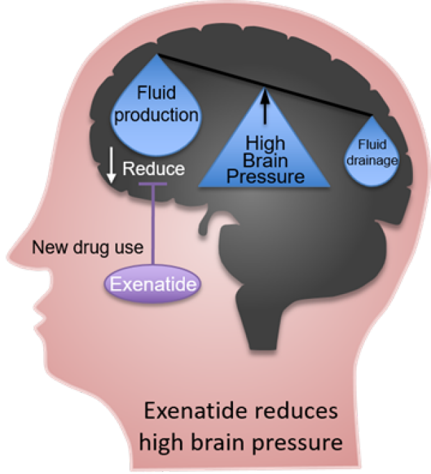
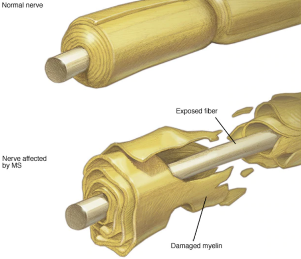
Some of the Neuro-Opthalmic concerns we investigate include Multiple Sclerosis, Optic Neuritis, IIH, and ICP.