No trials match your search ""
Our research team includes physicians, residents, medical students, research assistants, and volunteers. Our research topics include medical imaging, device validation, mobile application development, and pharmaceutical trials.
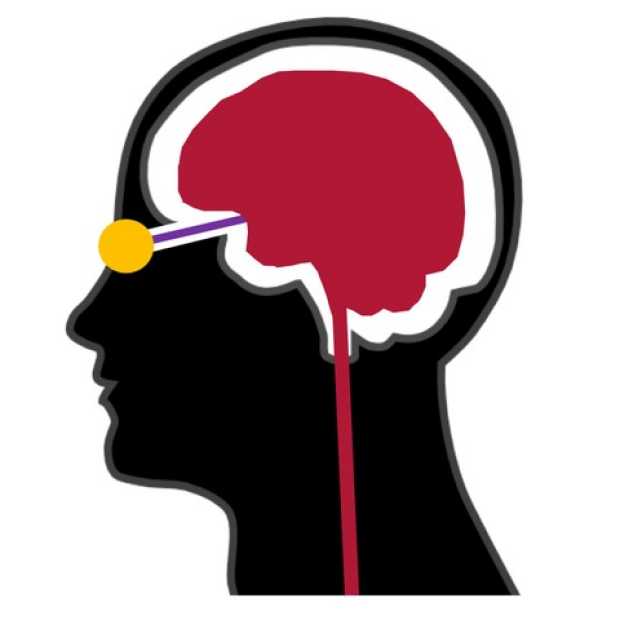
Some of the Neuro-Opthalmic concerns we investigate include Multiple Sclerosis, Optic Neuritis, IIH, and ICP.
No trials match your search ""
Other Trials that are accepting patients
Coming soon
In addition to the treatment trials described on this page we are actively studying IIH, vision in Multiple Sclerosis, and other conditions. See research projects (link) for more details.
Physiologically Based Markers of Idiopathic Intracranial Hypertension, Stanford University
Dr. Heather Moss
hemoss@stanford.edu
Permanent visual impairment due to papilledema, an optic neuropathy characterized by optic nerve swelling, occurs in approximately half of patients with IIH. There is a significant clinical need for non-invasive biomarkers that will advance diagnosis and management of IIH. The objective of my research is to establish physiologically based markers of retinal ganglion cell(RGC) function and retinal/cerebral vasculature as markers of IIH that detect abnormalities, monitor treatment and distinguish peripheral vision outcomes. I have demonstrated that retinal vein diameter changes over the course of disease. Through collaboration with Dr. Ali Alaraj, an endovascular neurosurgeon, we have defined characteristic changes in cerebral venous blood flow and pressure in IIH patients. Through collaboration with Dr. McAnany, a psychophysics expert, we have demonstrated alterations in objective markers of optic nerve function that correlate with other measures of disease in IIH patients. These results are laying the scientific and technical foundation for the development of these markers as clinical tools and clinical trial outcome measures. Furthermore, the results are advancing scientific understanding of the pathophysiology underlying papilledema and other optic neuropathies.
Risk factors for peri-operative vision loss, Stanford University, University of Illinois at Chicago
Dr. Heather Moss
hemoss@stanford.edu
Perioperative visual loss (POVL) is a devastating complication, with no known treatment or prevention, most commonly due to ischemic optic neuropathy (ION), and retinal arterial occlusion (RAO), and less commonly cortical blindness. We reported in 2009 that POVL had an estimated incidence of 3-10 cases/10,000 procedures in two of the highest volume surgical procedures.1 The resulting severe visual impairment costs > $27,000/y, or $675,000 during the estimated remainder of a middle-aged individual’s life from increased health care spending alone. Lost productivity costing > $250,000, and frequent litigation further increase costs. The emotional toll of sudden, unexpected visual loss is immeasurable. It is imperative to understand the risk factors for POVL in order to develop means to prevent these blinding complications. In collaboration with the University of Illinois at Chicago and University of Illinois at Chicago we are studying risk factors and developing a predictive model for perioperative visual loss (POVL) in spinal fusion and cardiac surgery.
Development of Portable Pupillometer
Megha Bindiganavale
mbindiga@stanford.edu
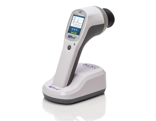
Pupil reactivity is an important clinical marker of optic nerve function. Quantitative pupillometry may offer advantages over the qualitative clinical pupil exam. Current commercial devices that provide this metric are cumbersome, expensive, and require operator expertise. Our goal is to configure and validate the use of a portable device to perform quantitative pupillometry. A portable pupillometer might be useful in a clinical and research setting for diagnosis and monitoring of optic neuropathies and neurological disease. Light stimulation and pupil recording protocols were implemented using the retEVAL commercial device and Lua programming language.
Modeling

Munam Wasi
munam@stanford.edu
Our goal is to build 3D geometric models of the eye from planar MRI imaging for finite element analysis. It is understood that one mechanism of disease/injury that translates from the brain to the eyes is high intracranial pressure, but it’s quite impossible to see what’s going on inside the head through conventional means, as post-mortem autopsies capture the eyes without forces acting upon them. MRI images allow us to take a snapshot in time and infer the forces acting upon an eye, in order to better understand disease progression and what actually defines normalcy. Ultimately we hope that these computational techniques can be married with other tools like OCT to build automated diagnostic systems and improve care through detection.
MS Study
Fareshta Khushzad
fkhush@stanford.edu
Our goal is to evaluate novel non-invasive eye imaging biomarkers, with a focus on blood vessels, and neurons, as potential surrogates of brain inflammation and degeneration. We hope to learn which quantitative retinal vessel metrics can be used to help characterize inflammation in patients with multiple sclerosis. The information is important as it will provide preliminary information to further investigate these metrics as biomarkers in future studies to determine retinal vessel markers as longitudinal markers of MS. This knowledge will eventually allow clinicians to easily and inexpensively characterize the inflammation in patients with MS to help preemptively treat patients and make better predictions on the prognosis of the disease.
ERG Study
Megh Patel, Fareshta Khushzad, Dr. Heather Moss
hemoss@stanford.edu
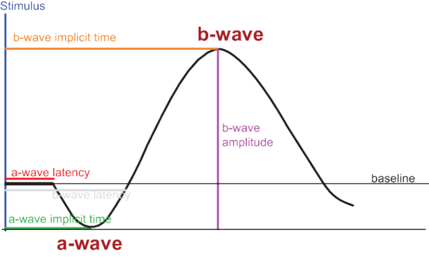
In clinical practice, diseases that affect the retina and optic nerve are monitored by subjective patient responses to visual stimuli. Electrophysiology tests provide objective markers of optic nerve and retina function that may compliment subjective measures of vision. This project aims to compare electrophysiological tests of optic nerve and retina function. We hope to learn if electrophysiological tests and pupil reactivity tests using different devices and different stimulation protocols are comparable and how they relate to subjective visual function measures.
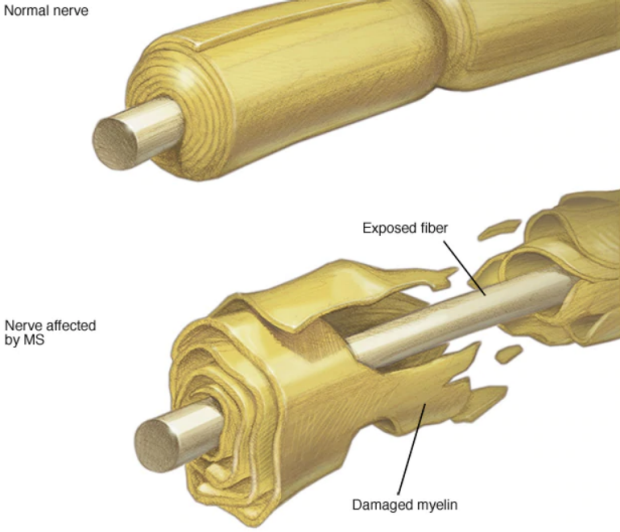
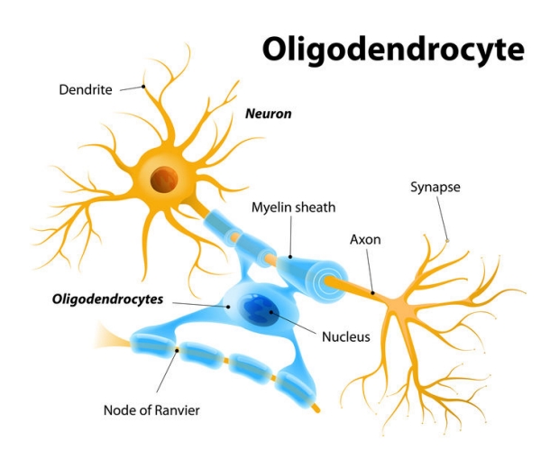
Comparison of Myelin Oligodendrocyte Glycoprotein Optic Neuritis Clinical Presentation between different ethnicities.
Tanyatuth Padungkiatsagul
tpadung@stanford.edu
During this past decade immunological markers of optic neuritis have been identified and became a substantial addition to patient care. Myelin Oligodendrocyte Glycoprotein, a novel serology marker, has linked to a certain variant of optic neuritis. Literatures from different part of the world reports some discrepancy in clinical presentation and prognosis. Ethnicity could be an important underlying factor for this variation, Thus we are investigating in our multi-center cohort of Myelin Oligodendrocyte Glycoprotein Optic Neuritis(MOGON) combined with published literatures in order to discover potential clinical variation among different ethnicity.