Heart Failure
Objective: MRI-based computational constitutive modeling presents a clinically viable myocardial (heart muscular tissue) stiffness estimation technique. Data necessary for this stiffness estimation include high-resolution cardiac geometry, diastolic kinematics, myocardial microstructural organization and boundary conditions. Using this MRI data and filling pressures, we can construct computational models to estimate myocardial stiffness. It is important to validate these methods in order to quantify the accuracy and precision of the stiffness estimates obtained. We have developed an experimental system incorporating 3D-printed hearts of myocardium-mimicking mechanical and MRI properties. These heart phantoms were embedded into an MRI-compatible setup to validate our MRI-based in vivo myocardial stiffness estimation approach. The approach is also being evaluated in heart failure patients.
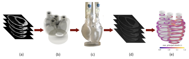
Figure 1: Stiffness framework validation (a) porcine MRI data; (b) geometric model; (c) manufactured heart phantom adapted with load ports; (d) heart phantom images; (e) computational modeling.
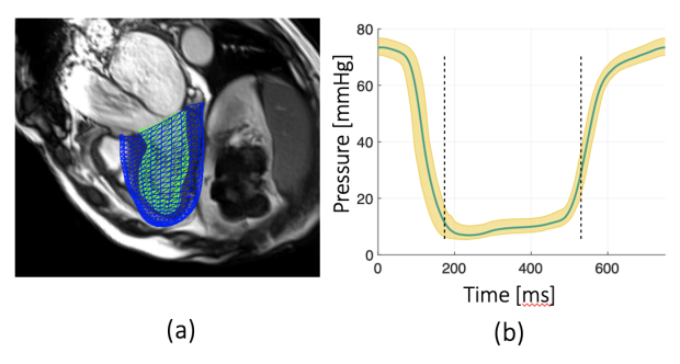
Figure 2: Myocardial stiffness estimation (a) HFpEF patient LV computational model generated from cine bSSFP MRI (b) Sample LV pressure used as boundary condition in computational model

Daniel B. Ennis, PhD
Professor Department of Radiology
Director of Radiology Research VA Palo Alto Healthcare System