Neuro Applications
Experimental study of intracranial hematoma detection with flat panel detector C-arm CT.
Arakawa H, Marks MP, Do HM, Bouley DM, Strobel N, Moore T, Fahrig R.
Department of Radiology, Stanford University Medical Center, 300 Pasteur Dr, Stanford, CA 94305-5105, USA.
BACKGROUND AND PURPOSE: Intracranial hemorrhage is a commonly acknowledged complication of interventional neuroradiology procedures, and the ability to image hemorrhage at the time of the procedure would be very beneficial. A new C-arm system with 3D functionality extends the capability of C-arm imaging to include soft-tissue applications by facilitating the detection of low-contrast objects. We evaluated its ability to detect small intracranial hematomas in a swine model.
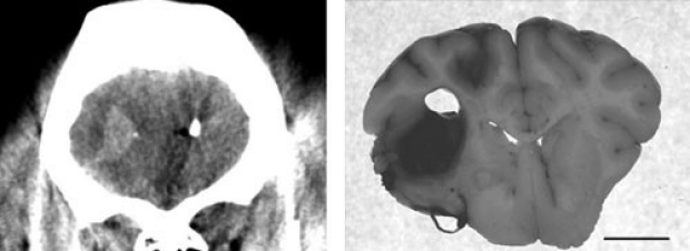
MATERIALS AND METHODS:Intracranial hematomas were created in 7 swine by autologous blood injection of various hematocrits (19%-37%) and volumes (1.5-5 mL). Four animals received intravascular contrast before obtaining autologous blood (group 1), and 3 did not (group 2). We scanned each animal by using the C-arm CT system, acquiring more than 500 images during a 20-second rotation through more than 200 degrees . Multiplanar reformatted images with isotropic resolution were reconstructed on the workstation by using product truncation, scatter, beam-hardening, and ring-artifact correction algorithms. The brains were harvested and sliced for hematoma measurement and compared with imaging findings.
RESULTS:Five intracranial hematomas were created in group 1 animals, and all were visualized. Six were created in group 2, and 3 were visualized. One nonvisualized hematoma was not confirmed at necropsy. All the others in both groups were confirmed. In group 1 (with contrast), small hematomas were detectable even when the hematocrit was 19%-20%. In group 2 (without contrast) C-arm CT was able to detect small hematomas (<1.0 cm(2)) created with hematocrits of 29%-37%. The area of hematoma measured from the C-arm CT data was, on average, within 15% of the area measured from harvested brain.
CONCLUSIONS: The image quality obtained with this implementation of C-arm CT was sufficient to detect experimentally created small intracranial hematomas. This capability should provide earlier detection of hemorrhagic complications that may occur during neurointerventional procedures.
Cerebral CT perfusion using an interventional C-arm imaging system: cerebral blood flow measurements.

Ganguly A, Fieselmann A, Marks M, Rosenberg J, Boese J, Deuerling-Zheng Y, Straka M, Zaharchuk G, Bammer R, Fahrig R.
Department of Radiology, Stanford University, California 94305-5488, USA.
BACKGROUND AND PURPOSE: CTP imaging in the interventional suite could reduce delays to the start of image-guided interventions and help determine the treatment progress and end point. However, C-arms rotate slower than clinical CT scanners, making CTP challenging. We developed a cerebral CTP protocol for C-arm CBCT and evaluated it in an animal study.
MATERIALS AND METHODS: Five anesthetized swine were imaged by using C-arm CBCT and conventional CT. The C-arm rotates in 4.3 seconds plus a 1.25-second turnaround, compared with 0.5 seconds for clinical CT. Each C-arm scan had 6 continuous bidirectional sweeps. Multiple scans each with a different delay to the start of an aortic arch iodinated contrast injection and a novel image reconstruction algorithm were used to increase temporal resolution. Three different scan sets (consisting of 6, 3, or 2 scans) and 3 injection protocols (3-mL/s 100%, 3-mL/s 67%, and 6-mL/s 50% contrast concentration) were studied. CBF maps for each scan set and injection were generated. The concordance and Pearson correlation coefficients (? and r) were calculated to determine the injection providing the best match between the following: the left and right hemispheres, and CT and C-arm CBCT.
RESULTS: The highest ? and r values (both 0.92) for the left and right hemispheres were obtained by using the 6-mL 50% iodinated contrast concentration injection. The same injection gave the best match for CT and C-arm CBCT for the 6-scan set (? = 0.77, r = 0.89). Some of the 3-scan and 2-scan protocols provided matches similar to those in CT.
CONCLUSIONS: This study demonstrated that C-arm CBCT can produce CBF maps that correlate well with those from CTP.