
zeego@Stanford Lab
System Description
The zeego system consists of a large area flat panel detector (30x40 cm) and a rotating anode x-ray tube mounted on a C-arm, which is then attached to a 6-axis robot. For this system, the C- mounting hardware was redesigned to reduce the overall weight of the component by more than 30%, providing enhanced stability and very accurate positioning control. The true strength of the system is its flexibility, making it an ideal system for a wide range of research applications. The specifics of the technology are described below, with a comparison against our current ceiling-mounted Artis dTA system. The system also has the standard advantages of all high-end C-arm systems, that real-time 2D high-frame rate high-spatial-resolution projection images and 3D CT images can be acquired using the same system. The specifications listed here are the latest available from the innovations group at Siemens AX in Forchheim, and have already been achieved in factory testing.
Hardware – The Robot and C-arm
KUKA is one of the world's leading manufacturers of industrial robots and automation systems for a variety of industries - from automotive and fabricated metals to food and plastics. The robot chosen for this system has six degrees of freedom, with an additional two degrees of freedom provided by rotation of the x-ray detector and x-ray tube to provide portrait and landscape acquisition modes (total angular range of 90°, tube and detector move in synchrony). The rotation center of the robot positioner can be placed anywhere in a sphere around the desired imaging volume (ie. variable isocenter), can be moved during acquisition, and the imaging volume itself can be easily relocated/moved. The robot can move at a maximum speed of 120°/s with both acceleration to maximum speed and deceleration to 0 requiring approximately 0.5s; motion accuracy and reproducibility has so far been verified for speeds up to 100°/s. This speed is twice that of the current Artis dTA and opens the door to a significant number of applications that require fast, serial volume CT images. The C-arm has a focal-spot-to-detector distance of 120 cm, and a focal-spot-to-isocenter of 78.5 cm if a standard 'propellor' rotational acquisition is performed. High-voltage, control and signal cables restrict total angular range of the system to 360° in propeller mode, and to ˜300° in sleeve mode.
Hardware – Digital Flat Panel Detector
The digital flat panel on this system is amorphous silicon with a CsI scintillator converter. It has a limiting resolution of 154μm, providing high spatial resolution of 3.25 lp/mm and good low-contrast resolution. The detector has been optimized for operation in the energy range of 70-100 kVp, with a 0.6-mm-thick CsI scintillator. Imaging chain hardware and software upgrades permit operation with a maximum frame rate of 80 fps in the 4x4 binning mode. The acquisition speeds and corresponding resolutions are summarized in Table A.I. All acquisitions are done in 14 bit gray scale resolution. Cubic interpolation of the images to a 1kx1k matrix is used only to fit the acquired images onto the 1024x1024 displays – all image data is stored at full resolution. The detector can function in 'overview' mode (zoom 0, diagonal 19") and has 5 additional zoom modes, with diagonals of 17" (zoom 1), 13" (zoom 2), 9", 6" and 4".
| Binning Mode | Pixel Size (μm) | Matrix Size | Max. Frame Rate (fps) | |
|---|---|---|---|---|
| Zoom 0 | unbinned | 154 | 2480x1920 | 7.5 |
| 2x2 | 308 | 1240x960 | 30 | |
| 4x4 | 616 | 616x480 | 60/80 | |
| Zoom 1 | unbinned | 154 | 1920x1920 | 7.5 |
| 2x2 | 308 | 960x960 | 30 | |
| 4x4 | 616 | 480x480 | 60/80 |
Table A.I Summary of pixel size, matrix size and associated frame rates that can be achieved with the 30x40 digital flat panel detector. The detector on the zeego will be capable of 80 fps.
Hardware - X-ray tube and Generator
The x-ray tube is the Megalix Cat125/15/40/80 three-focus high-performance x-ray tube assembly with metal center tube. The focal spot sizes are 0.3/0.6/1.0 mm with power ratings of 15/40/80 kW respectively, and operates in the range 50-125 kVp. The anode angle is 12°, providing sufficiently uniform irradiation over the large field of view of the flat panel. The x-ray tube normally operates in pulsed mode up to 60 pulses per second.
Operating Modes – Fluoroscopy
Digital pulsed fluoroscopy (low-dose imaging) is available at 10, 15 and 30 fps in 1k/12-bit matrix with real-time filtering for noise reduction including motion detection; low-dose options are available if lower frame rates can be tolerated, from 0.5-7.5 fps. Digital radiography and digital subtraction angiography is provided at frame rates ranging from 0.5-7.5 fps in original matrix size, full format and in zooms 1 and 2 in 2k matrix. High-speed acquisition for DSA, DR, and cardiac acquisition is provided at 7.5, 10, 15 and 30 fps, with acquisition and storage as indicated in Table A.I, and display in 1k matrix, 12-bit.
Operating Modes - 3D Imaging Acquisition
'Standard' Single-Volume 3D Image Acquisition: The zeego can rotate at a maximum speed of 120° per second, permitting the acquisition of a 'pi + fan angle' conebeam CT data set in under 2 s. Using the maximum frame rate of 120 fps, the limiting resolution of reconstructed volumes would be 592 μm/1.5 (magnification) ˜ 400 μm isotropic (ie. in-plane resolution is the same as slice thickness). The large 30x40 cm flat panel detector provides sufficient Z coverage (˜20 cm) to image the whole human brain, with a reconstructed in-plane FOV of diameter 26 cm. If slower rotations are acceptable (e.g. for high-resolution imaging of a static object) then imaging time can be extended in order to acquire sufficient numbers of high-resolution projection images around the object. The 2K image acquisition mode can be used with a slow, 20-s rotation to generate 3D volumes with isotropic voxel resolution of ˜ 100 μm.
'Multi-sweep' Multi-Volume 3D Image Acquisition: The Z-class group of C-arm imaging systems is the only C-arm-based imaging system that permits serial bi-directional sweeps to be acquired within a single acquisition. Turnaround time between 'forward' and 'backward' sweeps is on the order of 1s, and so with the zeego, 3D images can be acquired at a frame rate of ˜2.5 volumes/s. Maximum acquisition speed is matched to the maximum detector readout rate (80 fps), to provide 400 μm isotropic resolution in the reconstructed volumes. The data is stored after each sweep, and so in principle the number of serial sweeps that can be acquired in a single acquisition is limited only by the x-ray tube heat loading capacity and the total data storage capacity of the image acquisition system.
The multi-sweep capability has been combined with ECG-gating to provide reasonable temporal resolution in reconstructed high-resolution 3D images of moving objects with repeating motion such as the beating heart, and pulsatile vessels. Temporal resolution is optimized for a given heart rate by timing the start of each sweep such that maximum coverage over the cardiac cycle is achieved. The temporal resolution is therefore approximately limited by the number of sweeps and the heart rate of the subject. For example, for a heart rate of 60 beats per minute, if 6 sweeps are acquired during a single acquisition, then the temporal resolution will be ˜1s/6 = 166 ms. The total acquisition time for 6 sweeps is ˜15 s, within a reasonable breathhold.
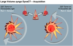
'Large Volume' Double-Sweep Acquisition: The 'large volume' data acquisition protocol acquires image data during a forward and backward sweep of the C-arm, with an offset in center of rotation between the two sweeps (see Figure A.2). Using new reconstruction algorithms, this effectively doubles the reconstructed FOV to 47 cm diameter with a z-extent of 18 cm. This reduces the impact of truncation (or missing projection data) for large objects that extend outside the standard 26-cm diameter FOV and increases the accuracy of reconstructed HU numbers. Alternatively, by rotating the x-ray detector by 90°, the whole thoracic spine could be imaged with a 35-cm diameter FOV and a z-extent of 25 cm. With the current factory-tested rotation speed of the zeego, such volumes can be acquired in ˜7s.
Reconstruction Software and Image Analysis
We highlight here some of the additional reconstruction and analysis packages available the zeego platform.
syngo InSpace 3D FlashRT is a high-end post processing workstation, with a Windows XP PC with syngo-based user software and network modules. The 3D volumes are reconstructed using standard FDK with a package of corrections implemented for truncation, beam hardening, non-stationary (but reproducible) center of rotation, and scatter.[22,23] Reconstructions can be completed very quickly : a 5s acquisition, 2x2 binning, 126 images requires only 20s, an 8s acquisition, 4x4 binning, 419 images requires 22s, and a 20s acquisition, 2x2 binning, 543 images requires 48s. This reconstruction speed is an order of magnitude faster than can be achieved using the current hardware and software in the Axiom lab.
syngo InSpace 3D/3D Fusion package provides spatial alignment and visualization of image data of a single patient where the image data has been generated at different points in time or by different imaging modalities. Fusion of morphological and functional information and therapy planning is supported.
syngo i-Pilot provides visualization and fading between live 2D fluoroscopic image and the matching 3D reconstruction, where the 'view' of the 3D volume can be modified according to zoom, SID and table position to match the fluoro image.
syngo iGuide provides integrated needle guidance for interventional procedures using definition of needle path on a DynaCT or external CT volume, definition of progression views for monitoring the needle insertion, and alignment and progression of the needle under fluoroscopic control with needle path overlaid on the live image of the acquisition system.
syngo InSpace EP is used for 3D visualization of the heart including automated segmentation of one or more ventricles/vessels of the heart, and especially provides optimized segmentation of the left atrium and pulmonary veins. The automatic segmentation works on preoperative 3D CT, MR or intraoperative 3D DynaCT Cardiac data.