Research
Examples of the many exciting projects in our group!

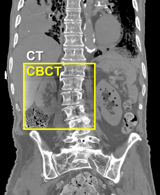
Zeego Lab
This robotic C-arm system is capable of fluoroscopic and CT imaging for anatomical, functional, quantitative, brain, cardiovascular, and body applications. Research projects include weight-bearing CT, dual energy imaging with fast kV switching, and imaging of lung airways.
Quantitative Imaging
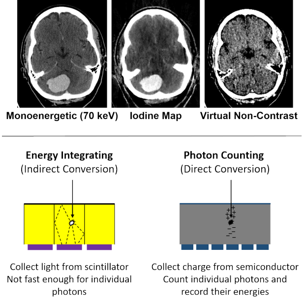
Material-specific quantification is performed by leveraging spectral information from methods like dual energy or photon counting imaging. Our group seeks to maximize the information obtained from these methods to enhance diagnostic and image-guidance capabilities. In particular, we are modeling the realistic behavior of photon counting detectors to better understand their performance.
Novel System Development
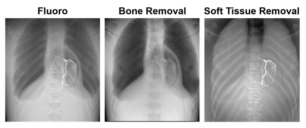
We continue to develop novel imaging systems, including new detector designs. We are evaluating a dual-layer flat-panel detector, which provides dual energy images with perfect spatial and temporal registration over a large area (43x43 cm2) and at high frame rates (up to 15 fps). We also have an NIH-funded project to investigate single-shot quantitative imaging (SSQI), which combines a primary modulator and a dual-layer detector to simultaneously measure scatter and dual energy with every image.
Artificial Intelligence
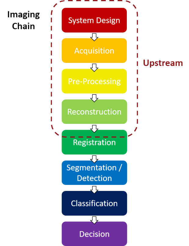
We are using AI, and particularly deep learning, to address various needs in CT. Our focus is on the "upstream" aspects of imaging, such as system design, acquisition, preprocessing, and reconstruction. In particular, we seek to maximize image quality while minimizing radiation dose to the patient.
Monte Carlo & Deterministic Transport
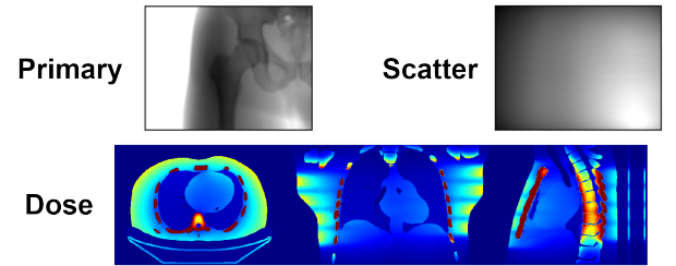
Photon transport can be modeled through stochastic Monte Carlo simulation or deterministic methods that solve the Linear Boltzmann Transport Equation. This enables us to determine scatter in an image and radiation dose in the object.
Model-Based Image Reconstruction
Our group develops advanced image reconstruction methods that incorporate knowledge of the imaging chain and physics, including statistical properties of the measurements. We also speed up the reconstruction via accelerated convergence algorithms and GPU implementation.