
Research
Ubiquitin Research
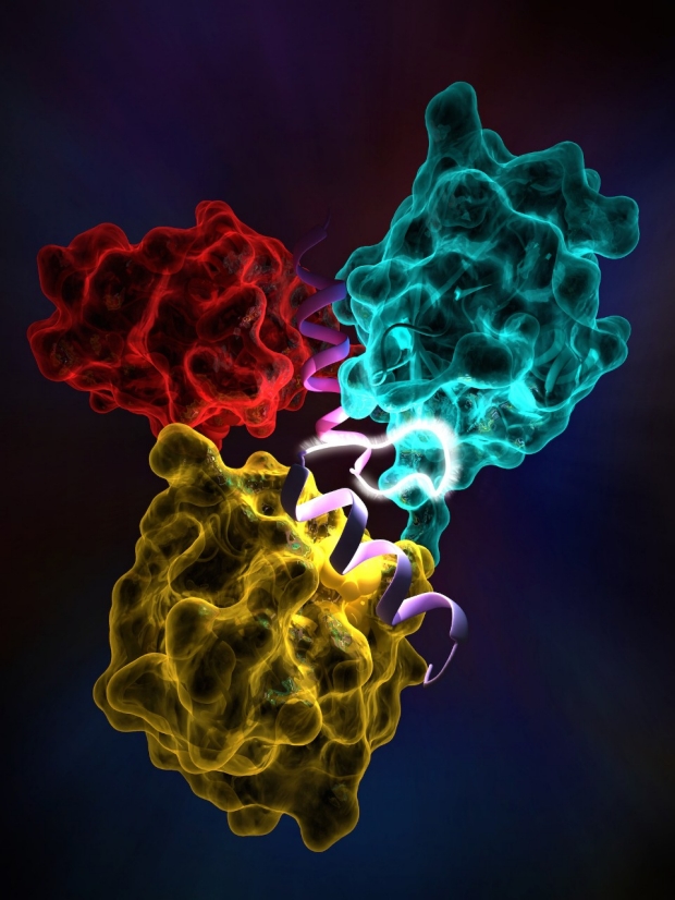
Ubiquitin is a small protein modifier that is ubiquitously produced in the cells and takes part in the regulation of a wide range of cellular activities such as gene transcription and protein turnover. The key to the diversity of the ubiquitin roles in cells is that it is capable of interacting with other cellular proteins either as a single molecule or as different types of chains. Ubiquitin chains are produced through polymerization of ubiquitin molecules via any of their seven internal lysine residues or the N-terminal methionine residue. Covalent interaction of ubiquitin with other proteins is known as ubiquitination which is carried out through an enzymatic cascade composed of the ubiquitin-activating (E1), ubiquitin-conjugating (E2), and ubiquitin ligase (E3) enzymes. The ubiquitin signals are decoded by the ubiquitin-binding domains (UBDs). These domains often specifically recognize and non-covalently bind to the different ubiquitin species, resulting in distinct signaling outcomes.
We apply a combination of the structural (including protein x-ray crystallography, small angle x-ray scattering, etc.) and biochemical techniques to study the ubiquitylation process by the E3 ubiquitin ligases and recognition of the ubiquitin chains by the proteins harboring ubiquitin-binding domains.
Illustration by Greg Stewart/SLAC

Photoenzyme Catalysis
Understanding the biological use of light to do chemical work remains one of the grand challenges of modern science. The overwhelming majority of the energy that fuels life and industry comes from biological photosynthesis, either current or historic (fossil fuel). Despite this, both the specific mechanism of photosynthesis and the general principles of how enzymes can employ light to do chemistry remain unknown.
With the ability of SLAC’s linac coherent light source (LCLS) to perform x-ray imaging at femtosecond timescales, the study of the mechanisms of enzyme-catalyzed photochemistry has formed a core component of the research conducted at the lab. This research was not possible before the LCLS made it possible to image biological catalysis at both atomic and femtosecond resolution. With this technology, direct measurement of the structure-dynamics-function behind biological photocatalysis have begun.
We are conducting time-resolved crystallography experiments to monitor structural changes in DNA photolyase as it performs a TT dimer repair reaction to address unknowns in the photolyase mechanism, and learn more about the general principles of enzyme-mediated catalysis. Our goal is a molecular movie of the photolyase reaction at Å/fs resolution.
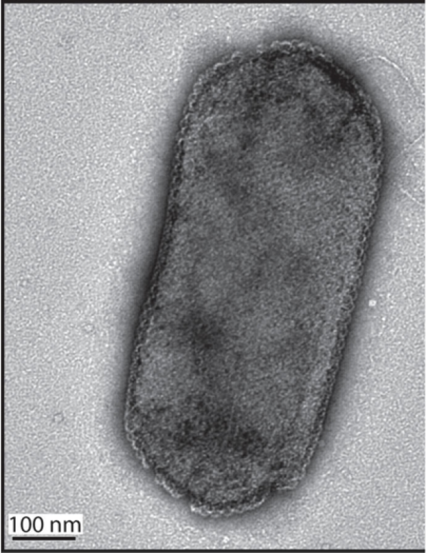
Nitrogen Cycling: ammonia oxidizing archaea
In collaboration with Chris Francis, Dept of Earth System Science, Stanford, and John Bargar, SLAC SSRL, we have recently initated a research program that leverages SLAC and Stanford strengths in x-ray structural molecular biology, biocomputation, and microbial ecology to deconstruct molecular-to-ecosystem structure-function relationships of nitrogen-cycling enzymes that drive global nitrogen (N) and carbon (C) turnover. This research program will advance our knowledge of the relationship between protein structure and function at the ecosystem scale by integrating three new, strategic efforts: (i) producing and characterizing environmental enzymes identified through metagenomic and metatranscriptomic approaches; (ii) examining protein structure at the scales of molecules and subcellular architecture; and (iii) using biocomputation, calibrated against biochemical assays and protein structures, to interrogate structure-function relationships encoded in large numbers of environmental metagenome sequences. Together, these approaches will offer for the first time the capability to systematically assess AOA enzyme structures (including both ammonia monooxygenase and nitrite reductase) produced under specific ecosystem conditions and the structural mechanisms that confer adaptive advantages. Such knowledge would transform our understanding of global biogeochemical N cycling and its molecular underpinning. Detailed information of environmental structure-function relationships could be used to help predict nutrient fluxes and land-atmosphere gas exchange. Structure-function knowledge will further provide important scientific insights, yet to be discovered, to engineer and utilize enzymes in biotechnological, environmental, and industrial applications.
Figure taken from Blainey et al., Plos One 2011, 6, (2), DOI 10.1371/journal.pone.0016626.
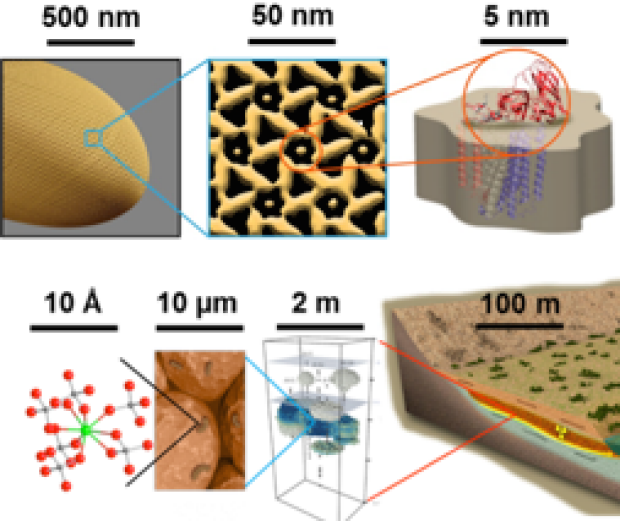
Multiscale imaging
Multimodal, multiscale imaging modalities will be developed and integrated to understand how molecular level events of key enzymes and protein network are connected to cellular and multi-cellular functions through intra-cellular organization and interactions of the key machineries in the cell. Cryo electron tomography will be employed to investigate subcellular organization of of surface layer proteins and key enzyme complexes at several nm resolution. Higher resolution structures of the key enzymes and S-layer proteins will be determined using SSRL and LCLS X-ray facilities as well as a cryo electron microscope to be installed at SLAC late 2016 to early 2017 at SLAC. Larger scale organization of these proteins will be studied by solution X-ray scattering while electronic states and interplay between the key enzymes including ammonia monooxygenase and nitrite reductases will be studied by X-ray spectroscopy (absorption and emission). Their spatio-temporal arrangements in the cell membrane will be further studied by X-ray fluorescence imaging using resonance of Cu in the key enzymes, and correlated with cryoEM and super-resolution optical microscopy.