Research
Molecular studies of human sarcomas and carcinomas
In our laboratory we study human tumors with the ultimate goal of developing new diagnostic markers, new biomarkers for prognosis and response to treatment and to develop novel therapeutic targets. We use and develop a range of techniques that focus on extracting genomic and proteomic data from patient derived archival material (formalin-fixed paraffin-embedded (FFPE).
Genomic analyses include RNAseq, exome seq and 3SEQ for gene expression profiling and mutation calling. 3SEQ (3’-end sequencing for expression quantification) is a novel procedure that we developed for gene expression profiling from FFPE tissue using next-generation sequencing. Tissue microarrays generated with FFPE material from thousands of tumor samples are analyzed with immunohistochemistry and in situ hybridization techniques to validate findings from the molecular studies. We now have over 500,000 digital images from these TMA’s stored in our image database, TMAD.
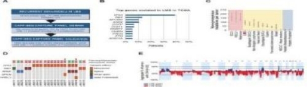
Leiomyosarcoma
We are using gene expression profiling and comparative genomic hybridization to determine genes that are involved in the malignant transformation of smooth muscle cells in a wide variety of locations. In our most recent work we confirmed the existence of molecular subtypes in LMS. Future studies will investigate the clinical relevance of these findings, specifically the sensitivity of each subtype to current clinical treatment modalities and therapies that are under development, such as the CD47-based approach. In collaboration with the laboratories of Drs. Ash Alizadeh and Max Diehn at Stanford, we are studying circulating tumor DNA in LMS patients. The goal is to use the levels of circulating LMS DNA as a measurement for tumor in the patient and to evaluate the response to therapy.
Our work on LMS work is supported by donations from Leiomyosarcoma Direct Research, the National Leiomyosarcoma Foundation and the Liddy Shriver Initiative.
Gastrointestinal stromal tumors
While clinically relevant GIST is extremely rare, small (< 1cm) GIST are quite common. We are performing a study that tries to identify the molecular changes in “mini-GISTs” to clinically significant tumors.
Desmoid type fibromatosis
We are studying desmoid type fibromatosis, using gene expression profiling and tissue microarray analysis with the intent of identifying diagnostic markers for this tumor and to finding novel targets for therapy.
Macrophages in Human tumors
Macrophages constitute a major subset of cells in the cancer tumor microenvironment (TME), but despite decades of research in murine models and in vitro studies on human blood monocyte-derived macrophages there are no reliable markers to detect the various macrophage functions in the human TME and no systematic study on human macrophage in vivo functional diversity has been performed. A convergence of three novel technologies (Smart-3SEQ, CIBERSORTx and MIBI) now offers an opportunity to discover novel markers by studying macrophages in vivo at the microscopic level in the actual TME rather than in a model thereof. Central to our approach is a novel technology, Smart-3SEQ (developed by the West laboratory), that allows gene expression profiling on groups of cells or even individual cells isolated by laser capture microdissection from formalin fixed paraffin embedded material where numerous samples with known clinical outcome are readily available. This allows for a physical purification of histologically identified cells in specific locations to provide exact quantification of GEP from cells within distinct locations within the TME. An orthogonal approach, that allows cross validation with the LCM data, is offered by a novel bioinformatics approach (CIBERSORTx, developed by the Newman laboratory) that offers an “in silico purification” of immune cell-specific mRNA levels using FFPE-derived GEP data. A unique multi-marker imaging technique “Multiplexed ion beam imaging” (MIBI, developed by the Angelo laboratory) will be used to characterize new macrophage markers in the context of the other constituents of the TME, including blood vessels, fibroblasts, and T- and B-lymphocytes subsets.

Combination approach for detecting different types of alterations in circulating tumor DNA in leiomyosarcoma. Clin Cancer Res. 2018 Jun
Mutational landscape of LMS and design of LMS-specific CAPP-Seq selector
