Research
Middle Ear Imaging Project
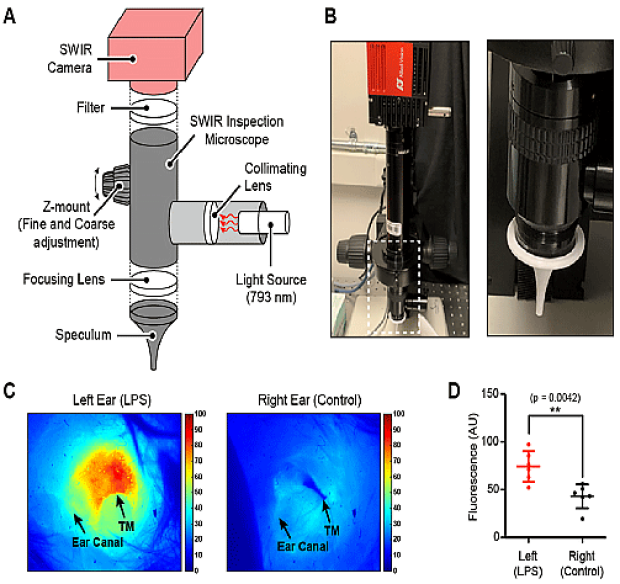
Otitis media (OM, inflammation in the middle ear) is one of the most common illnesses among children under five years of age. It is known to affect about 90% of children worldwide. Accurate diagnosis and treatment of OM is critical as it can lead to additional complications such as speech and language development delays, brain abscesses, meningitis, and even hearing loss. We have developed a medical otoscope for SWIR imaging. Due to its higher wavelengths (1-2 microns), SWIR light can penetrate deeper in the tissue compared to the visible light pneumatic otoscopy, enabling a better view of anatomy behind the tympanic membrane and higher contrast to middle ear fluid due to increase water absorption in the SWIR.
In collaboration with Bogyo lab at Stanford, we have developed smart fluorescent probes to increase the contrast of infectious OM in SWIR imagining. Our aim is to distinguish between non infected (otitis media with effusion) and infected fluid (acute otitis media) by combining SWIR otoscopy with inflammation targeted probes.
Cholesteatoma Imaging
Cholesteatoma is a proliferation of abnormal skin that can develop in the middle ear and can erode and destroy important structures associated with hearing and balance. If left untreated in can lead to potentially life-threatening complications such as meningitis and brain abscesses. Surgical removal is the only alternative and often requires multiple procedures due to a high recurrence rate. We are developing imaging techniques to better identify cholesteatoma in a surgical field to prevent recurrence and understand the molecular changes that precede its formation.
The Traveling Pediatric Simulators

We have developed pediatric specific surgical simulators to teach new generations of doctors around the world. We are firm believers that surgical training is a gradual process and basic techniques and surgical fundamentals can be learned in surgical models prior to being performed in patients.

We look both at commonly performed procedures to see how they can be performed in a safer and more efficient way to minimize anesthesia exposure and to those rare conditions where it is difficult to gain adequate experience during training.
Our simulators have been around the world: Europe, Asia, North America, South America looking for partners to put us on the map in Africa, and penguins in Antarctica (Pittsburgh penguins need not apply).
Treatment of Middle-Ear Effusions
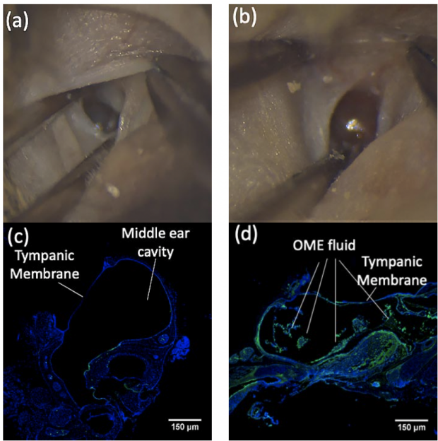
OM with effusion (OME) is characterized by the presence of middle ear fluid containing thick secretions behind the tympanic membrane (i.e., ear drum). There is no effective medicine for treating OME. Current clinical practice relies on “mechanical” removal of secretions by surgery via ventilation tube placement, which requires general anesthesia and carries the risk of a tympanic perforation. Our goal is to develop an alternative treatment, to reach the secretions and alter their rheological properties to facilitate their drainage from throat/nose.
Cisplatin Ototoxicity and Prevention
Cisplatin is one of the most widely used chemotherapeutic agents, but one of its major side effects is an irreversible sensorineural hearing loss, which occurs in 50-70% of patients with cancer treated with cisplatin. Most of the existing diagnostic approaches rely on indirectly measuring effects and morphological changes, which might not be a direct reflection of the amount of cisplatin accumulated. The purpose of this study is to develop a label-free and objective approach for real-time assessment of cisplatin accumulation in the inner ear. Successful implementation of the work would lead to a new method for identifying cisplatin-induced ototoxicity.