Measuring and Understanding the Ankle Brachial Index (ABI)
The Ankle Brachial Index (ABI Test) is an important way to diagnose peripheral vascular disease. The index compares the systolic blood pressures of the arms and legs to give a ratio that can suggest various severity of peripheral vascular disease.
Introduction to Measuring the Ankle Brachial Index
The Ankle Brachial Index (ABI) is the systolic pressure at the ankle, divided by the systolic pressure at the arm. It has been shown to be a specific and sensitive metric for the diagnosis of Peripheral Arterial Disease (PAD). Additionally, the ABI has been shown to predict mortality and adverse cardiovascular events independent of traditional CV risk factors. The major cardiovascular societies advise measuring an ABI in every smoker over 50 years old, every diabetic over 50, and all patients over 70.
Measuring the Ankle Brachial Index
Venous Testing Using A Doppler
Technique for Measuring the Ankle Brachial Index
The ABI is performed by measuring the systolic blood pressure from both brachial arteries and from both the dorsalis pedis and posterior tibial arteries after the patient has been at rest in the supine position for 10 minutes. The systolic pressures are recorded with a handheld 5- or 10-mHz Doppler instrument. Usually a standard blood pressure cuff can be used at the ankle. As with arm pressures, the most accurate pressure readings are obtained when the blood pressure cuff is appropriately sized to the patient's lower calf (immediately above the ankle). It is recommended to begin with the right arm, then the right leg, then the left leg, and finally the left arm, as the blood pressure may drift during the exam, and the two arm pressures at the beginning and end of the exam provide for some quality control.
Measuring the brachial pressure
- The patient should be in the supine position. Place the blood pressure cuff on the arm, with the limb at the level of the heart. Place the ultrasound gel in the antecubital fossa over the patient's brachial pulse. Place the transducer of the handheld Doppler on the gel, and position the transducer to maximize the intensity of the signal. Inflate the cuff to about 20 mmHg above the expected systolic blood pressure of the patient. The Doppler signal should disappear. Then slowly deflate the cuff, approximately 1 mmHg/sec. When the Doppler signal re-appears, the pressure of the cuff is equal to the brachial systolic pressure. Record the brachial systolic pressure.
Measuring the ankle pressures
- Place the cuff immediately proximal to the malleoli. Place ultrasound gel on the skin overlying the dorsalis pedis (DP) and posterior tibial (PT) arteries in the foot. The Doppler signal of the DP can often be found slightly lateral to the midline of the dorsum of the foot. Using a standard hand-held Doppler probe and the ultrasound gel, locate the signal from the DP. Slowly move the Doppler until the strongest signal is heard. To measure the systolic pressure at the DP artery, inflate the cuff until you no longer hear the signal. Then slowly deflate using the same technique used in the arms until the Doppler signal re-appears. Record the measurement.
- Next, measure the systolic pressure of the PT artery. The PT signal is detected posterior to the medial malleolus. Once again, using the Doppler with ultrasound gel, locate the signal, and follow the process described above to measure the PT systolic pressure. Repeat both measurements on the opposite leg.
Calculating the ABI
- An ABI is calculated for each leg. The ABI value is determined by taking the higher pressure of the 2 arteries at the ankle, divided by the brachial arterial systolic pressure. In calculating the ABI, the higher of the two brachial systolic pressure measurements is used. In normal individuals, there should be a minimal (less than 10 mm Hg) interarm systolic pressure gradient during a routine examination. A consistent difference in pressure between the arms greater than 10mmHg is suggestive of (and greater than 20mmHg is diagnostic of) subclavian or axillary arterial stenosis, which may be observed in individuals at risk for atherosclerosis.
- Calculated ABI values should be recorded to 2 decimal places.

.gif)
While measurement of the ankle brachial index in an important diagnostic tool, it is important to remember other signs of peripheral vascular disease. These include history and exam findings such as pain with walking (claudication), paraesthesia (numbness), paralysis (weakness), pulselessness (of dorsalis pedis and posterior tibial pulsus) and pallor of distal extremities. Both paralysis and paraesthesia are often seen in very severe ischemia to the legs.
.gif)
Interpreting the Ankle Brachial Index
- Normal ABI ranges from 1.0 — 1.4
- Pressure is normally higher in the ankle than the arm.
- Values above 1.4 suggest a noncompressible calcified vessel.
- In diabetic or elderly patients, the limb vessels may be fibrotic or calcified. In this case, the vessel may be resistant to collapse by the blood pressure cuff, and a signal may be heard at high cuff pressures. The persistence of a signal at a high pressure in these individuals results in an artifactually elevated blood pressure value.
- An value below 0.9 is considered diagnostic of PAD.
- Values less than 0.5 suggests severe PAD.
- Individuals with such severe disease may not have sufficient blood flow to heal a fracture or surgical wound; they should be considered for revascularization if they have a non-healing ulcer.
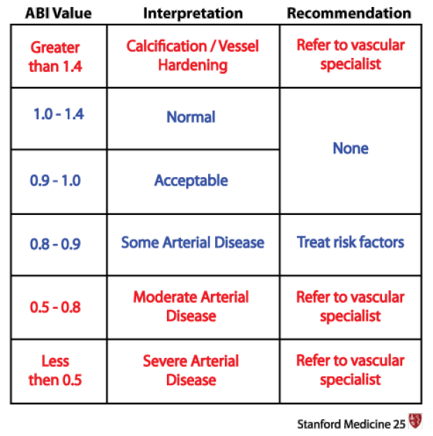
| ABI Value | Interpretation | Recommendation |
| Greater than 1.4 | Calcification/Vessel Hardening |
Refer to vascular specialist |
| 1.0-1.4 | Normal | None |
| 0.9-1.0 | Acceptable | None |
| 0.8-0.9 | Some Arterial Disease |
Treat risk factors |
| 0.5-0.8 | Moderate Arterial Disease |
Refer to vascular specialist |
| Less than 0.5 |
Severe Arterial Disease |
Refer to vascular specialist |
ABI Value |
Interpretation |
Recommendation |
|---|---|---|
| Greater than 1.4 | Calcification/Vessel Hardening |
Refer to vascular specialist |
| 1.0-1.4 | Normal | None |
| 0.9-1.0 | Acceptable | None |
| 0.8-0.9 | Some Arterial Disease |
Treat risk factors |
| 0.5-0.8 | Moderate Arterial Disease |
Refer to vascular specialist |
| Less than 0.5 |
Severe Arterial Disease |
Refer to vascular specialist |
Key Learning Points
- Learn the technique of measuring the ankle brachial index and interpreting the results
- Learn other uses of the Doppler probe for venous testing
Related to Ankle Brachial Index Exam
The Stanford Medicine 25
- Aortic Regurgitation Exam
- Ankle Brachial Index
- Ankle and Foot Exam
- Ascites & Venous Patterns
- Bedside Ultrasound
- Breast Exam
- Cardiac Second Sounds
- Carpal Tunnel Exam
- Cerebellar Exam
- Deep Tendon Reflexes
- Dermatology Exam: Acne vs. Rosacea
- Dermatology Exam: Learning the Language
- Dermatology Exam: Nevi (Mole) Exam
- Fundoscopic Exam (Ophthalmoscopy)
- Gait Abnormalities
- Hand Exam
- Hip Region Exam
- Internal Capsule Stroke
- Involuntary Movements and Tremor Diagnosis: Types, Causes, and Examples
- Knee Exam
- Liver Exam
- Low Back Exam
- Lymph Node Exam
- Neck Vein Exam
- Pelvic Exam
- Precordial Movements in the Cardiac Exam
- Pulmonary Exam: Percussion & Inspection
- Pupillary Responses
- Pulsus Paradoxus and Blood Pressure Measurement Techniques
- Rectal Exam
- Spleen Exam
- Tarsal Tunnel Exam
- Thyroid Exam
- Tongue Exam
- Liver Disease, Head to Foot
- Visit the 25
- Shoulder Exam Tutorial
- Parkinson's Disease Exam
- Diastolic Murmurs Exam
- Dermatology Exam: Nevi (Mole) Exam