Fevers, mild confusion and this retinal finding… Diagnosis?

March 24, 2016
A 42 year old female presents to Stanford hospital with fevers, chills and mild confusion. You perform a fundoscopic examination and see this (image below). What is the diagnosis?
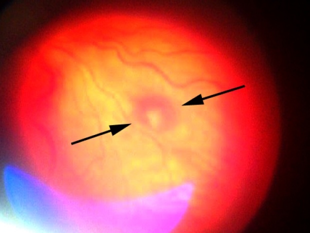
Answer: Roth spot
What is a Roth Spot?
A Roth spot, seen most commonly in acute bacterial endocarditis is a red spot (caused by hemorrhage) with a characteristic pale white center. This white center usually represents fibrin-platelet plugs (1).

Is a Roth Spot pathognomonic for bacterial endocarditis?
No. This is a common misconception. A Roth spot can be can be seen in leukemia, diabetes, intracranial hemorrhage, hypertensive retinopathy, subacute bacterial endocarditis and in HIV retinopathy (1,2).
What is the history of this name?
“Roth spot” derives from Moritz Roth, a Swiss pathologist who described retinal white spots and retinal red spots in 1872. Of note, he never described the presence of a retinal red spot combined with the white center spot. It was actually described by Mortiz Litten (yes, same first name) 6 years later who would coin the term “Roth spot” we still use today. Therefore, some prefer to call this finding a Litten spot orLitten sign. Mortiz Litten was a German physician known for describing vitreous bleeding seen in subarachnoid hemorrhage. He was son-in-law to Ludwig Traube, known for Traube’s space (learn more here) and Traube’s sign (pistol shot systolic sound heard over the femoral artery in aortic regurgitation).
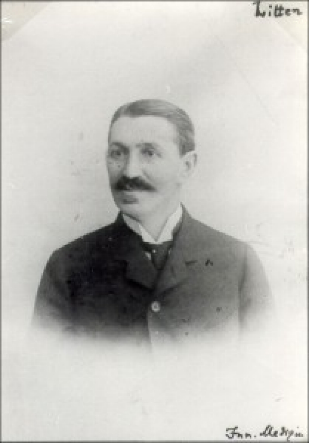
Dr. Moritz Litten
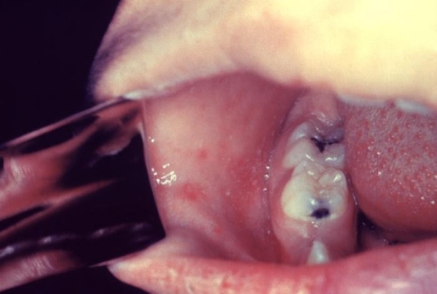
What’s a Koplik spot and why it mentioned here?
Koplik spots are seen in measles, usually 1-2 days before the rash appears spreading from head to toe. Koplik spots are seen in the mucosa of the mouth. Like Roth spots, Koplik spots are often described as a pale white center on a red background. Because of this, we sometimes refer to Roth spots as Koplik spots of the eye (though the cause and pathogenesis of each lesion differs). However, Koplik spots are usually many in number and not lesions have a red background.
What other physical exam findings should you look for with suspected endocarditis?
A careful cardiac examination is very important. Listen for murmurs (such as new diastolic murmur from valve damage leading to aortic regurgitation) and gallops.
Some findings include:
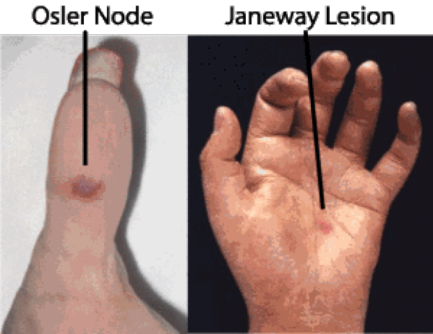
Remember to check both the hands and feet!
What happened to the patient?
She received IV nafcillin at Stanford hospital and eventually recovered. Just one day after observing the Roth spot, it was markedly faded and eventually gone in days. She fully recovered from the endocarditis after an extended course of antibiotics.
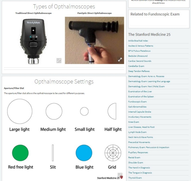
How was the image of the Roth spot taken?
This image was taken at the bedside of this patient at Stanford hospital using a Welch-Allyn PanOptic and iphone 4. The patient’s pupils were not dilated in this case.
Learn more on the technique of the fundoscopic exam:
Visit our newest video on fundoscopy found here and our Stanford Medicine 25 page on the fundoscopic exam to learn the techniques of fundoscopy and pathology of the retina. All clinicians should have confidence in visualizing the retina. The fundoscopic exam is the only time you can see arteries and is an opportunity to aid in many diagnosis of the body!
Source/References:
- Fred HL. Little black bags, ophthalmoscopy, and the Roth spot. Tex Heart Inst J. 2013;40(2):115-6.
- Van Uitert RL, Solomon GE. White-centered retinal hemorrhages: a sign of intracranial hemorrhage. Neurology 1979;29 (2):236–9.
- Image credit: Dr. Moritz Litten
- Image credit: Koplik spots